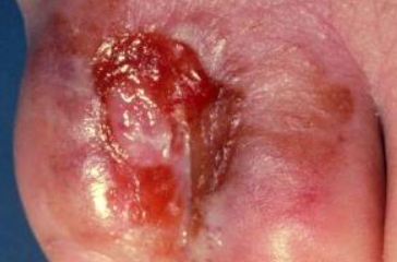
Tinea pedis (Athlete’s foot) - fungal infections on the skin of the feet. ICD-10 codes: B35.3
The most common causative agents of tinea pedis are the pathogenic fungi Trichophyton rubrum (90%) and Trichophyton mentagrophytes var. interdigitale. Less commonly, these diseases are caused by Epidermophyton floccosum and Candida species.
Infection with pathogenic fungi can occur through direct contact with an infected person, as well as through footwear, clothing, personal items (bathroom mats, sponges, nail care tools, etc.), visiting gyms, saunas, swimming pools. Skin abrasions, tears in the interdigital folds caused by friction, increased sweating or dryness of the skin, inadequate drying after water procedures, narrow interdigital spaces, vascular diseases in limbs, and other factors can facilitate fungal penetration of the skin.
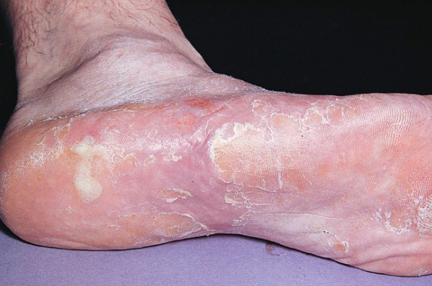
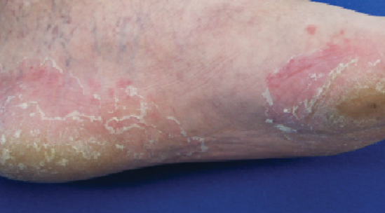
Mycoses can become widespread, especially in the presence of coexisting conditions such as endocrine disorders, often diabetes, immune disorders, genodermatoses, blood disorders, and the use of antibiotics, corticosteroids, and cytostatic drugs.Squamous hyperkeratotic type of tinea pedis (Moccasin-variety)
Intertriginous type of Tinea Pedis
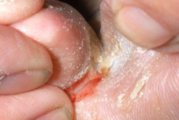
This form is characterized by the appearance of macerations and fissures in the interdigital folds, often between the III and IV, IV and V toes.
In the interdigital folds, a well-defined area of macerated and eroded skin forms against a background of swelling and redness. Eruptions in the form of erosions and deep cracks, along with wetness, are often painful and accompanied by itching and burning. In some cases, blister-like eruptions may also be observed. Subjectively, there is itching, burning, and tenderness in the affected areas.Vesiculobullous type of tinea pedis
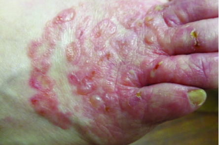
It manifests as numerous vesicles with a thick covering located on the arch and lower lateral surfaces of the foot, as well as in the interdigital folds. The predominant localization is on the arches of the feet. The eruptions may involve extensive areas of the soles as well as the interdigital folds and the skin of the toes. They coalesce to form large multichambered vesicles that rupture to produce moist, pink erosions. The blisters are usually located on unaffected skin. As the inflammation progresses, the skin becomes erythematous and swollen, resembling acute dyshidrotic eczema. It is subjectively characterized by pruritus.
It typically follows a chronic, intermittent course with exacerbations in spring and fall. Exacerbations are accompanied by severe pruritus. The dyshidrotic form is often caused by T. mentagrophytes var. interdigitale.Ulcerative type of tinea pedis
Tinea pedis in children
In children, involvement of the feet is characterized by fine scaling on the inner surface of the terminal phalanges of the toes, often in the 3rd and 4th interdigital folds or under the toes, accompanied by redness and maceration. The skin on the soles may be either unchanged or have an increased skin pattern. This condition is associated with pruritus. Exudative forms of the disease are more common in children than in adults and may involve the hands as well as the feet.
Diagnosis of dermatophytosis is based on clinical presentation and laboratory findings:
Microscopic examination of scales from affected skin areas. Cultural and PCR methods are used to determine fungal species.
If systemic antifungals are prescribed, a biochemical blood test is recommended to assess bilirubin, AST, ALT, GGT, alkaline phosphatase and glucose levels. For treatment-resistant forms of onychomycosis, ultrasound examination of superficial and deep vessels is recommended.- Palmoplantar Psoriasis
- Contact Dermatitis
- Juvenile plantar dermatosis
- Pitted Keratolysis
- Interdigital Erythrasma
- Hyperkeratotic Eczema
- DyshidroticEczema
- Candidal Intertrigo
- Keratolysis Exfoliativa
- Climacteric Keratoderma
- Secondary Syphilis
Treatment Goals:
- Clinical resolution
- Negative results in microscopy test
Topical Therapy
Topical antifungals:
- Isoconazole cream, applied 1-2 times daily for 4 weeks, or
- Terbinafine spray or dermgel, applied 2 times daily until clinical manifestations resolve, or
- Ciclopirox cream, applied 2 times daily until clinical manifestations resolve, or
- Clotrimazole cream, ointment, or solution, applied 2 times daily until clinical manifestations resolve, or
- Ketoconazole cream or ointment, applied 1-2 times daily until clinical manifestations resolve, or
- Bifonazole cream, applied 1-2 times daily for 5 weeks.
- Econazole cream, applied 2 times daily until clinical manifestations resolve, or
- Miconazole cream, applied 2 times daily until clinical manifestations resolve, or
For significant hyperkeratosis in tinea, a preliminary removal of the epidermal stratum corneum is performed using bifonazole, applied once daily for 3-4 days.
Systemic therapy
If topical therapy is ineffective, systemic antifungals are prescribed:
- Itraconazole 200 mg orally daily after meals for 7 days, then 100 mg orally daily after meals for 1-2 weeks, or
- Terbinafine 250 mg orally daily after meals for 3-4 weeks, or
- Fluconazole 150 mg orally after meals once a week for at least 3-4 weeks.
Prevention
Primary prevention: Foot care aimed at preventing microtraumas, abrasions, elimination of hyperhidrosis (aluminum chlorohydrate 15% + decylene glycol 1%) or dryness of the skin (tetranyl U 1.5% + urea 10%), flat feet, and others.
Secondary prevention: Disinfectant treatment of footwear, gloves once a month until complete healing:
Chlorhexidine bigluconate, 1% solution.
