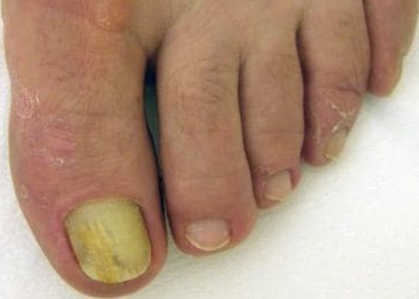
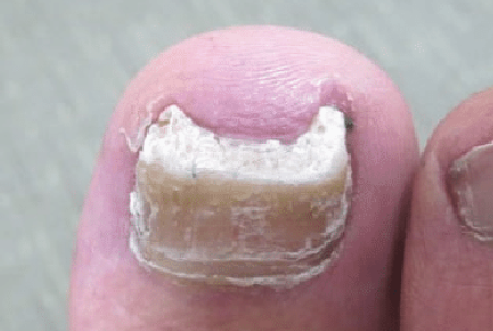
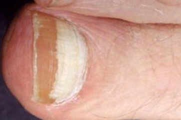
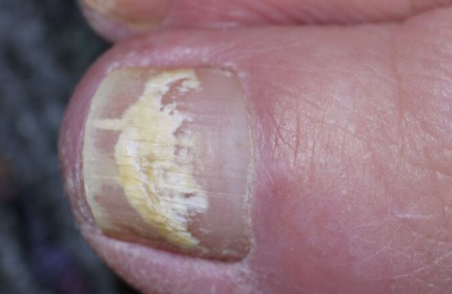
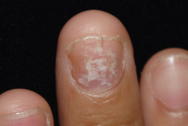
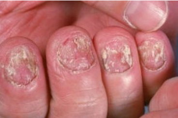
Onychomycosis is a fungal infection of the nail. ICD-10 code: B35.1.
Fungal nail infections affect 15-20% of the population between the ages of 40 and 60. The prevalence increases with age and infections are lifelong with no spontaneous remission.
Trauma from wearing tight shoes predisposes to infection. Dermatophytes are responsible for nearly 90% of onychomycosis cases, with the most common pathogens being Trichophyton rubrum and Trichophyton mentagrophytes. Other species such as Aspergillus, Cephalosporium, Fusarium, and Scopulariopsis, often considered contaminants or non-pathogenic, may also infect the nail plate. Multiple pathogens may be present on a single nail.
Once established, a fungal infection will persist. It can interfere with social interactions and cause pain if the nail plate becomes deformed. Thickened nail plates and underlying hyperkeratotic masses can be uncomfortable to wear shoes.Fungal nail infections may be associated with tinea manuum and pedis or may occur independently. All skin and nail surfaces must be examined to rule out other conditions that may mimic onychomycosis. Dermatophytes cause four distinct clinical forms of onychomycosis based on the type of fungal invasion of the skin.
Distal subungual onychomycosis
Proximal subungual onychomycosis
White superficial onychomycosis
Endonyx Onychomycosis
Candidal onychomycosis
There is a tendency to label any process that affects the nail plate as a fungal infection; however, many other skin conditions also can alter nail structure. Approximately 50% of thickened nails are not infected with fungi.
Subungual hyperkeratotic masses and nail plates are examined with KOH for the presence of fungal hyphae.
Fungal species are identified prior to prescribing systemic antifungal medications. Fungal culture is performed to determine the presence of dermatophytes-organisms that are susceptible to itraconazole, terbinafine, and fluconazole. Clear guidelines for monitoring patients receiving terbinafine, itraconazole, or fluconazole are lacking. A reasonable approach would include compete blood count and liver function tests before and 6 weeks after treatment. Laboratory monitoring is not required in patients receiving pulse therapy with itraconazole.
Histologic examination of nail material is performed when potassium hydroxide test and culture testing are negative but clinical suspicion remains high.- Nail psoriasis
- Leukonychia
- Onycholysis
- Yellow nail syndrome
- Subungual warts
- Onychopapilloma
The choice of treatment depends on the clinical form of onychomycosis and the number of nails involved.
Topical treatment:
For topical antifungal treatment to penetrate the nail plate, a specific vehicle is required to deliver the drug through the nail plate.
Currently, the following delivery systems are available in most European countries: nail lacquers containing amorolfine 5% and ciclopirox 8%. Amorolfine lacquers are applied weekly, while ciclopirox is applied daily. Nail lacquers are effective as monotherapy for the treatment of superficial white onychomycosis and distal subungual onychomycosis limited to the distal surface of the nail with involvement of a few fingers. Treatment should be continued for 6-12 months. Nail lacquers are also used in severe onychomycosis in combination with systemic antifungals to reduce treatment duration and increase cure rates. Daily application of nail lacquer may also prevent recurrence of onychomycosis after successful treatment.
Systemic Treatment
Distal subungual onychomycosis that has spread to the proximal nail, endonyx onychomycosis, and proximal subungual onychomycosis require systemic therapy. Systemic treatment with terbinafine or itraconazole achieves mycological cure in over 90% of fingernail infections and approximately 80% of toenail infections. Recurrence rates are approximately 15-20% after one year. Treatment efficacy may be increased by combining systemic therapy with topical nail lacquer treatment.
Terbinafine is administered at a dose of 250 mg/day for 6 weeks for fingernail infections and 12 weeks for toenail infections, or as pulse therapy at 500 mg/day for 1 week per month. Terbinafine has the highest cure rate and longest remission. It is ineffective against certain Candida species. Itraconazole is used as a pulse therapy at a dose of 400 mg/day for 1 week per month. The duration of treatment is 2-3 months for fingernail infections and 3-4 months for toenail infections. Data on the efficacy of fluconazole in the treatment of onychomycosis are recent and further research is needed to determine the optimal dosage and duration of treatment. The approved dose of fluconazole is 150 mg once weekly, but higher doses (300 mg once weekly) are likely to be more effective. Treatment should be continued for at least 6 months. Griseofulvin can be effective at very high doses when given for several months, but other medications are more effective. Patients are monitored after 6 weeks and at the end of systemic therapy. Infected nail plates are debrided at each visit. In most cases, nails do not clear within 12 weeks. Patients should be informed that the medication will remain in the nail plate for months and will continue to eliminate fungi.
Chemical or surgical nail removal may be combined with systemic antifungal agents to remove localized fungal masses in cases of total onychomycosis.
Sequential treatment with itraconazole and terbinafine has been used to increase cure rates. A suggested regimen includes two courses of itraconazole at 400 mg/day for 1 week per month, followed by one or two courses of terbinafine at 500 mg/day for 1 week per month. Assessment of clinical response takes several months due to slow nail growth. Recurrence and reinfection are not uncommon (up to 20% of cured patients). Prevention includes regular application of nail lacquer to previously affected nails and use of topical antifungal agents on the soles and interdigital spaces.
