Pityriasis lichenoides occurs equally in individuals of all races, ethnic groups, and geographic regions. It is more common in males than in females (2:1 ratio). Pityriasis lichenoides occurs predominantly in individuals between the ages of 15 and 30, and rarely in children and the elderly. Chronic forms of the disease are diagnosed three times more frequently than acute forms.
Etiology is unknown. It is believed that the development of pityriasis lichenoides is associated with the formation of a hypersensitivity reaction to various infectious agents. Some authors consider this condition to be a self-limiting lymphoproliferative skin disease.
In acute pityriasis lichenoides varioliformis acuta, CD30+ marker expression is found on lymphocytes in the eruptive areas. In chronic cases, there is partial loss of the CD7+ marker, which is characteristic of cutaneous lymphoma.
Classification:
- L41.0 Pityriasis Lichenoides et Varioliformis Acuta (Mucha-Habermann Disease, PLEVA).
- L41.1 Pityriasis Lichenoides Chronica (PLC).
Pityriasis Lichenoides et Varioliformis Acuta
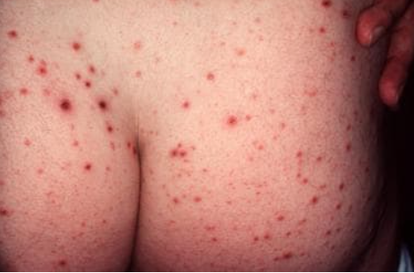
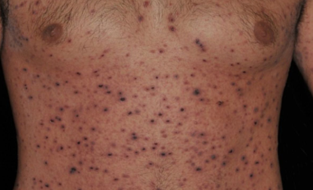
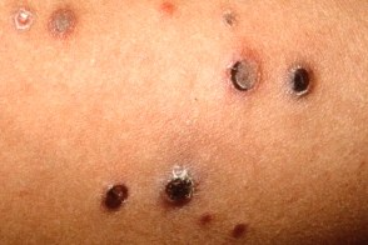
This condition develops rapidly, typically within a few days to 1-2 weeks. It is characterized by a true polymorphism of the eruption, with the simultaneous appearance of papules, pustules, papulovesicles and hemorrhagic macules. Due to the waves of eruptions, there is an evolutionary polymorphism of elements: pustule or papule - erosion or ulcer - crust - secondary spot. Pathognomonic for the acute form of pityriasis lichenoides are papules with a hemorrhagic crust in the center, leaving small scars after resolution, and varioliform elements - pustular vesicles with a central dimple. Skin involvement is widespread and symmetrical.
Most often the eruptions are localized on the chest, back, abdomen and flexural surfaces of the proximal parts of the limbs. The skin of the palms, soles, hairy areas of the head, and face are unaffected.
In most cases, the general condition is unaffected, but sometimes fever, lymphadenopathy, general weakness, and pruritus may occur.
Duration is usually unpredictable and the disease can last from several weeks or months to even years. However, acute pityriasis lichenoides varioliformis usually has a shorter duration than the chronic form. In some cases, acute form may progress to chronic form.Ulcerative-Necrotic Pityriasis Lichenoides et Varioliformis Acuta (Mucha-Habermann Disease)
Ulcerative-Necrotic Pityriasis Lichenoides et Varioliformis Acuta (Mucha-Habermann Disease): Characterized by an acute onset, development of febrile temperature (38-39°C), chills, generalized weakness and malaise, abdominal pain, headache, and lymphadenopathy. Single or, more commonly, multiple papules and/or plaques 5-15 mm in diameter appear on the skin. Rapid necrosis develops in their centers, followed by the formation of painful ulcers. Granulation of the ulcers leads to the formation of scars.
Primary Diagnostic Criteria:
- Fever
- Acute eruption of generalized ulcerative-necrotic papules and plaques.
- Rapid and progressive course without any tendency toward spontaneous resolution.
- Histopathological features as in pityriasis lichenoides et varioliformis acuta.
Additional Diagnostic Criteria:
- History of acute Pityriasis Lichenoides and Varioliformis in the patient's medical history.
- Involvement of oral mucosa.
Pityriasis Lichenoides Chronica
This condition is characterized by a slow onset (weeks to months), a prolonged course (several years), and alternating periods of exacerbation and remission. Subjective sensations are usually absent, although some patients may experience mild pruritus.
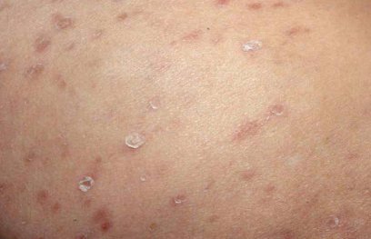
Primary morphologic element is a flat, round papule measuring 4-10 mm. It is initially pink or red in color and then turns yellowish-brown. The infiltrate at the base of the papule is minimal and the eruptions do not coalesce or cluster. Eruptions are typically located on the chest, abdomen, back, proximal parts of the limbs, and are particularly characteristic on the inner surface of the shoulders. Eruptions are not observed on the face, scalp, palms and soles.
Papules are firm with a smooth surface. When scraped, they show scaling (symptom of hidden scaling) followed by pinpoint bleeding (symptom of purpura). As papules develop, they take on a brownish color. Approximately 1-2 weeks later, a tightly adherent, centrally located, and well-demarcated serous brown scale appears on their surface, which, when carefully removed, completely detaches (scale sign). After complete resolution of the papule, a matte white scale remains in its place as a thin film attached to the underlying skin. Eruptions may occur at different stages during the course of the disease.
After several weeks, papules spontaneously resolve, leaving behind secondary hyperpigmented or hypopigmented spots. There is usually a tendency for improvement during summer months, probably due to natural ultraviolet radiation.Pityriasis Lichenoides et Varioliformis Acuta:
- Chickenpox
- Lymphomatoid papulosis
- Transient Acantholytic Dermatosis (Grover's Disease)
- Papular Syphilid
- Polymorphous Angiitis
- Eczema herpeticum
Pityriasis Lichenoides Chronica:
- Psoriasis
- Lichen planus
- Gianotti-Crosti Syndrome
- Papular Syphilid
- Pityriasis Rosea
Topical treatment:
Emollients are prescribed to restore the skin's water-lipid balance, retain moisture, enrich the skin with lipids, and reduce subjective sensation.
High-potency topical glucocorticosteroids are recommended in repeated courses every 2-3 months:
- Alclometasone dipropionate cream applied twice daily to the affected areas for 1-2 weeks.
- Betamethasone cream or ointment applied twice daily to the affected areas for 1-2 weeks.
- Mometasone furoate cream or ointment applied twice daily to the affected areas for 1-2 weeks.
- Methylprednisolone aceponate cream or ointment applied twice daily to the affected areas for 1-2 weeks.
Systemic treatment
Chronic Pityriasis Lichenoides:
- In cases of a resistant course, retinoids are prescribed: Acitretin, 25-50 mg per day orally for 6-8 weeks.
Pityriasis Lichenoides et Varioliformis Acuta:
- Antibiotics:
- Clarithromycin, 0.5 g orally twice daily for 10-14 days or
- Doxycycline, 0.1 g orally twice daily for 10-14 days.
- Erythromycin, 200 mg five times a day for 10-14 days.
- Glucocorticosteroids: Prednisone, 20-60 mg per day orally (2/3 of the daily dose after breakfast, 1/3 after lunch) with gradual reduction to complete discontinuation over 6-8 weeks.
- Methotrexate, 10-25 mg per week orally for 6-8 weeks.
- Cyclosporine A, 2.5-4.0 mg per kg of body weight orally per day for 6-8 weeks.
- Dapsone, 50-100 mg per day orally for 4-6 weeks.
- Retinoids: Acitretin, 25-50 mg per day orally for 6-8 weeks.
- UV Therapy: For chronic and acute forms of lichenoid parapsoriasis, effective therapies include non-selective UV therapy (UVA + UVB), selective UV therapy (UVB), and narrowband UVB (311 nm).


