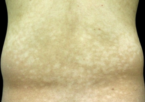
Progressive macular hypomelanosis is a skin condition caused by subtypes of Propionibacterium acne and characterized by the appearance of hypopigmented macules on the trunk and limbs. The ICD-10 code for this condition is L81.9.
The prevalence of the condition is difficult to estimate because it is often mistaken for pityriasis versicolor or vitiligo. The disease occurs between the ages of 13 and 45 (typically 20-26), with a higher prevalence in females than males (7:1 ratio). It predominantly affects individuals with skin types III-VI.
It is currently thought to be caused by infection with certain subtypes (probably type III) of Propionibacterium acne, which are assumed to produce substances that inhibit melanogenesis and/or melanosome distribution. Evidence supporting this theory includes:
- Detection of bacteria in smears and scrapings from the affected areas during microbiological examination.
- Red fluorescence of follicles under Wood's lamp in the affected areas due to the production of porphyrin by Propionibacterium acne.
- Rapid response to topical antibiotics.
However, it is unclear why the prevalence of the disease is not higher in individuals with acne, where Propionibacterium acne plays an important role in the pathogenesis. Isolated familial cases suggest a genetic predisposition to the disease, while onset and progression during pregnancy are attributed to hormonal factors.
The disease begins with the appearance of faint round hypopigmented macules on the abdomen and lower back, which rapidly progress and spread over the trunk, including the back and buttocks, merging into large hypopigmented macules of various shapes. The surface of the macules is smooth and without scaling. The eruptions are symmetrical, and involvement of the extremities and neck is rare, with no involvement of the scalp. There have been isolated cases of macules appearing on the face. There are no subjective symptoms. The disease is chronic and in most cases the rash spontaneously resolves after several years (typically 3-5 years). There is no apparent association with systemic or malignant diseases.Diagnosis is based on clinical presentation and history. A potassium hydroxide (KOH) test and culture are used to rule out fungal infection, while the red-colored perifollicular fluorescence under Wood's lamp in the affected areas confirms the diagnosis.
Histologic findings are nonspecific and typically show a normal number of melanocytes with decreased epidermal melanin content compared to healthy skin.- Pityriasis versicolor
- Pityriasis alba
- Vitiligo
- Post-inflammatory hypopigmentation
- Hypopigmented form of mycosis fungoides
- Leprosy
- Sarcoidosis
- Secondary syphilis
- Clear cell papulosis
- Pityriasis lichenoides
- Idiopathic guttate hypomelanosis
- Arsenical hypomelanosis
- Eruptive hypomelanosis
- Bier spots
- Visceral leishmaniasis (kala-azar)
- Combination of 1% clindamycin and 5% benzoyl peroxide gel once daily + UV phototherapy for 20 minutes per day, 3 times a week.
- Psoralen + PUVA therapy.
- Narrowband UV-B therapy at 311 nm, 1-3 times per week for 12 weeks.
- Oral doxycycline 100 mg daily for 6 weeks + insolation.
- Oral isotretinoin 10 mg daily for 2-4 months.

