Seborrheic dermatitis is a chronic recurrent skin condition associated with increased secretion of sebum, changes in its qualitative composition, and characterized by localization in areas rich in sebaceous glands - on the scalp, face, upper part of the trunk, and folds. ICD-10 code: L21.
The prevalence of seborrheic dermatitis worldwide is 1-3% in adults and 50-70% in infants in the first 3 months of life. Males are more affected than females in all age groups (3.0% vs. 2.6%), suggesting that the condition may be related to sex hormones such as androgens. There are two age peaks of seborrheic dermatitis - in childhood (first few months of life) and at 40-70 years old.
The etiology and pathogenesis are unclear. The development of the disease is facilitated by the activation of the lipophilic yeast Malassezia spp., increased secretion of sebum and changes in its qualitative composition associated with psychosocial stress, stressful situations, hormonal, immune and neuroendocrine disorders, and the use of certain medications.
The condition is frequently observed in HIV-infected individuals (30% - 83%), mostly in HIV patients with a CD4+ T-lymphocyte count between 200 and 500/mm³, and a decrease in the CD4+ count is often associated with exacerbations of the disease. These observations suggest that immunologic defects may play a role in the pathogenesis of the disease.
Seborrheic dermatitis is also associated with neurological and psychiatric disorders, including Parkinson's disease, neuroleptic-induced parkinsonism, tardive dyskinesia, head trauma, epilepsy, facial paralysis, spinal cord injury and depression. Associations have been reported with chronic pancreatitis in alcoholics, hepatitis C virus, and genetic disorders such as Down syndrome.
In addition, seborrheic dermatitis of the face may occur in patients receiving PUVA therapy. There is evidence that eruptions mimicking seborrheic dermatitis may occur in zinc and nicotinic acid deficiency.
Classification:
- L21 Seborrheic dermatitis
- L21.0 Seborrhea of the scalp
- L21.1 Infantile seborrhoeic dermatitis
- L21.8 Other seborrheic dermatitis
- L21.9 Seborrheic dermatitis, unspecified
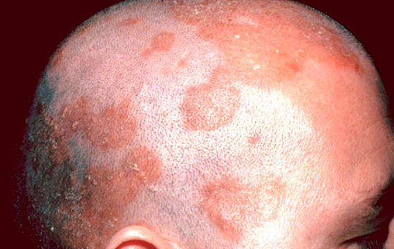
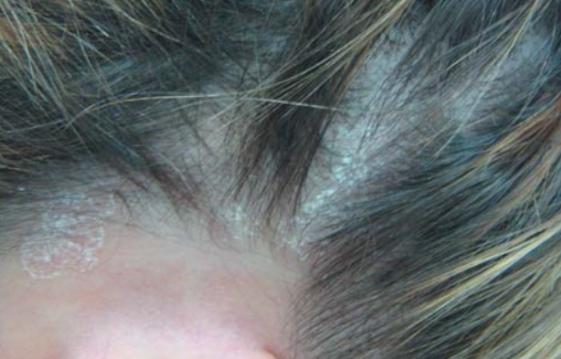
Scalp Seborrheic Dermatitis
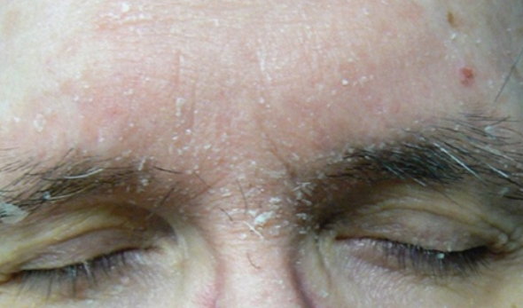
Seborrheic Dermatitis of the Face
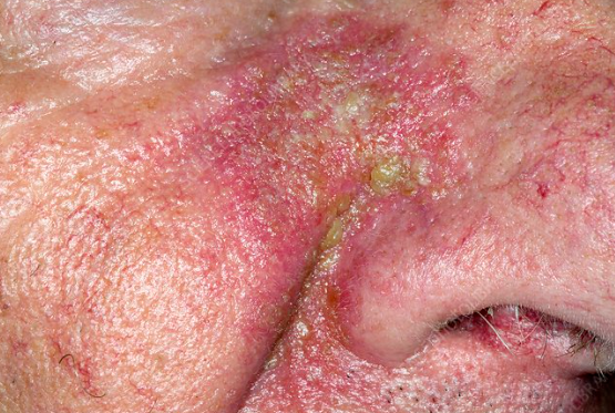
Perinasal Seborrheic Dermatitis
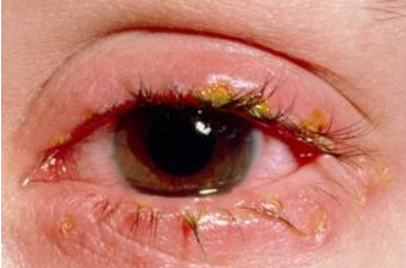
Seborrheic Blepharitis
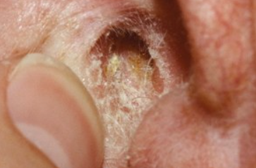
Ear Seborrheic Dermatitis
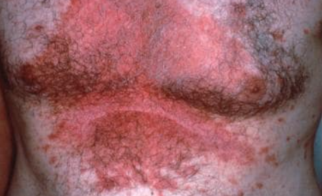
Chest Seborrheic dermatitis
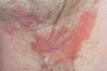
Intertriginous Seborrheic Dermatitis
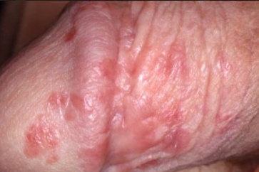
Intertriginous areas such as behind the ears, neck, axillary and groin folds, under the breasts and around the belly button are affected.
Erythema, swelling, and painful cracks often appear behind the ears and on the skin under the earlobes. In the axillary folds, eruptions occur in the center and gradually spread to the surrounding skin. Both folds are affected. Clinically, they appear as scaly erythema or as merging crusts and cracks. Seborrheic dermatitis in the axillary folds bears a strong resemblance to allergic dermatitis from deodorants but differs from dermatitis caused by clothing items (the latter is localized at the periphery of the fold and spares the center). Seborrheic dermatitis of the inguinal and perianal folds closely resembles dermatomycosis or candidiasis and sometimes presents with inverse psoriasis. The combination of psoriasis and seborrheic dermatitis (sebopsoriasis) in these folds is not uncommon. Erythema, swelling, oozing, painful cracking, and scaly crusts often develop in skin folds.Anogenital Seborrheic Dermatitis
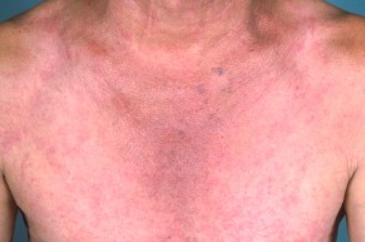
Generalized Seborrheic Dermatitis
It is characterized by a widespread process affecting multiple areas of the body. In adult patients, generalized seborrheic dermatitis is often associated with lymphadenopathies and may resemble a fungal infection (tinea).
Seborrheic dermatitis may be associated with or exacerbated by certain systemic diseases. Parkinson's disease is often associated with severe refractory seborrheic dermatitis of the face and scalp. Severe skin changes resembling seborrheic dermatitis may complicate diabetes mellitus, especially in obesity, celiac disease, malnutrition, epilepsy, and drug-induced skin conditions (especially when treated with neuroleptics such as haloperidol, gold preparations, or arsenic).Cradle Cap (Crŭsta Lacʹtea)
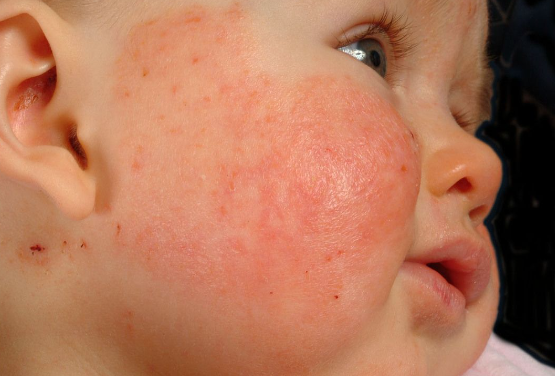
Seborrheic dermatitis in infants
Leiner disease
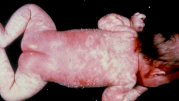
The coalescence of foci leading to erythroderma is described as Leiner disease and is characterized by three main symptoms: generalized rash in the form of erythroderma with desquamation, diarrhea, and hypochromic anemia. Delayed development and absence of neutrophil chemotactic factor C5a are noted.
The disease typically develops immediately after birth, less frequently in individuals older than 1 month of age, with erythematous infiltrated foci of bright red color and abundant desquamation, mainly in skin folds (groin, axillary). In the perineal area, on the buttocks and in the skin folds, the skin becomes macerated, cracked and may ooze.
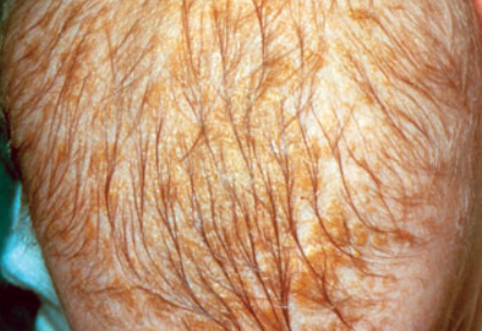
Greasy yellow scales on the scalp (cradle cap) spread to the eyebrow area and earlobes.
The overall condition of affected individuals is severe. Skin involvement is combined with gastrointestinal disturbances (frequent loose stools, vomiting), weight loss, hypotrophy, widespread candidal infection, and various infectious diseases. Without treatment, children develop severe toxic-septic conditions.Adults:
- Psoriasis
- Pemphigus erythematosus
- Pemphigus foliaceus
- Allergic contact dermatitis
- Tinea faciei
- Tinea corporis, capitis
- Lupus Erythematosus
- Perioral Dermatitis
- Rosacea
- Acneiform Dermatoses
- Pityriasis Rosea
- Secondary Syphilis
- Large plaque Parapsoriasis
Infants:
- Atopic Dermatitis
- Scabies
- Langerhans Cell Histiocytosis
- Psoriasis
- Neonatal Lupus Erythematosus
General Remarks on Therapy
- The choice of treatment strategy depends on the severity of clinical manifestations, the duration of the disease, and the effectiveness of previously administered therapies.
- The condition requires regular treatment using systemic and topical therapies over an extended period.
- For topical treatment, medications with anti-inflammatory, antipruritic, antifungal properties are used.
- In acute stage with severe itching and sleep disturbances, antihistamines and sedatives are recommended.
Treatment Regimens
Topical Treatment
Topical Glucocorticosteroids
- Betamethasone valerate 0.1%, cream, ointment, applied once daily for 7-14 days, or
- Betamethasone dipropionate 0.025%, cream, ointment, applied once daily for 7-14 days, or
- Hydrocortisone butyrate 0.1%, cream, ointment, applied twice daily for 7-14 days, or
- Methylprednisolone aceponate 0.1%, cream, ointment, applied once daily for 7-14 days, or
- Mometasone furoate 0.1%, cream, ointment, applied once daily for 7-14 days.
Topical calcineurin inhibitors
- Tacrolimus 0.03%, 0.1% ointment, twice a day for up to 6 weeks, maintenance therapy - twice a week as needed.
- Pimecrolimus 1% cream, twice a day for up to 6 weeks, maintenance therapy - twice a week as needed.
Topical antifungals
Ketoconazole, bifonazole, and ciclopiroxolamine creams and shampoos may be used for treatment. Prophylactic use of ketoconazole helps maintain remission. Bifonazole and ciclopiroxolamine may be prescribed in shampoo form three times a week. The shampoo should be applied to the scalp and beard area. The application time is 5-10 minutes before rinsing. After remission is achieved, the frequency of shampoo use may be reduced to twice a week or as needed.
Subsequent treatment involves using low-potency topical glucocorticosteroids and pastes containing 2-3% tar, naphthalan oil, or 0.5-1% sulfur.
Systemic Therapy
For severe itching, antihistamines are recommended:
- Acrivastine 8 mg orally twice daily for 14-20 days, or
- Loratadine 10 mg orally once daily for 10-20 days, or
- Cetirizine 5 mg orally twice daily for 10-20 days.
- Fexofenadine 120-180 mg orally once daily for 10-20 days, or
Tactics in Case of Treatment Failure
In severe cases or resistance to topical therapy, oral antifungal drugs may be prescribed.
- Itraconazole 200 mg orally once daily for the first week of treatment, then 200 mg orally once daily for the first 2 days of each subsequent 2-11 months of treatment, or
- Terbinafine 250 mg orally once daily continuously for 4-6 weeks or 12 days per month continuously for 3 months, or
- Fluconazole 50 mg orally once daily for 2 weeks or 200-300 mg once weekly for 2-4 weeks, or
- Ketoconazole 200 mg orally once daily for 4 weeks.

