Steroid atrophy is the most common side effect of the use of topical corticosteroids. Code ICD-10 as L90.9
It is most commonly observed in children and young women and occurs as a result of prolonged and regular use of topical steroids on the skin. The risk of developing atrophy depends on the class of corticosteroid used, the frequency and duration of application, and the site of application of the medication. It is more likely to occur with the use of fluorinated preparations. Atrophy may be observed as early as two weeks after initiation of therapy with very high-potency topical steroids, especially when applied under an occlusive dressing, and within four weeks when less potent preparations are used.
The pathogenesis of atrophy is caused by the following effects of steroids:
- Epidermal keratinocyte proliferation inhibition.
- Inhibition of collagen synthesis (types 1 and 3) in the dermis.
- Inhibition of fibroblasts and hyaluronan synthase 3 enzyme, leading to reduced hyaluronic acid in the extracellular matrix, resulting in skin atrophy.
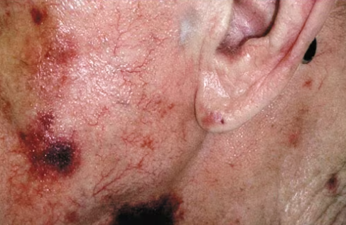
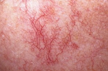
Stimulation by steroids of nitric oxide release from endothelial cells of dermal blood vessels, leading to abnormal capillary dilation and the development of telangiectasias in the affected area.
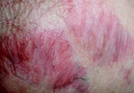
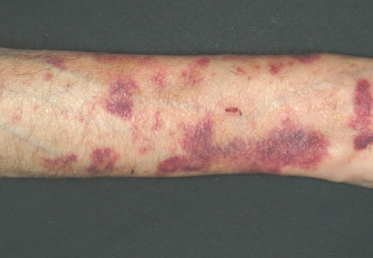
Steroid-induced protein degradation results in the loss of intercellular substances, causing blood vessels to lose their surrounding dermal matrix and become fragile, leading to the appearance of purpura and ecchymoses at the site of trauma.
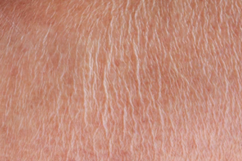
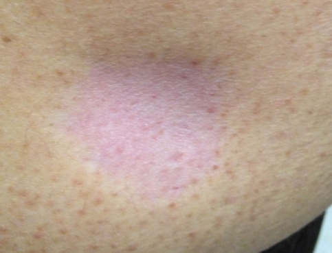
Steroids also inhibit melanin synthesis and disrupt the normal function of melanocytes, which can lead to hypopigmentation or, less commonly, hyperpigmentation in the affected areas.Superficial (epidermal) atrophy:
Dermal (deep) atrophy:
Pits:
Telangiectasia:
Purpura:
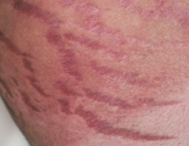
Stretch marks (striae):
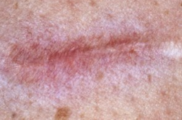
Pseudoscars:
- Stretch marks from physical stretching (obesity, pregnancy, physical exertion).
- Radiation dermatitis.
- Chronic sun exposure.
- Scar tissue from burns.
- Lipodystrophy from insulin injections.
- Morphea.
- Lichen sclerosus.
- Atrophie blanche in stasis dermatitis.
Epidermal atrophy and pigmentary changes are reversible after discontinuation of the drug. Resolution of skin lesions may take several months to several years.
Dermal atrophy is irreversible.
Treatment includes the use of topical retinoids (0.1% tretinoin cream). It is recommended that sunscreen be used on sun-exposed areas, as exposure to ultraviolet light may worsen the atrophy.

