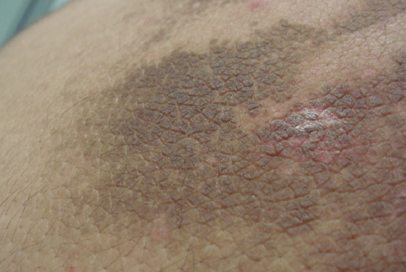
Terra firma forme dermatosis (also known as Duncan dirty dermatosis) is a chronic benign dermatosis of unknown etiology characterized by the presence of hyperpigmented plaques resembling unwashed dirt. These plaques are resistant to regular washing with soap and water, but are easily removed from the skin surface with 70% isopropyl alcohol. ICD-10 Code: L85.9
The condition can occur at any age, but is more common in children. The prevalence is unknown. The etiology and pathogenesis of the disease are unknown. It tends to be more prevalent during warm seasons. It is hypothesized that there is an unknown disorder and retention of keratinization leading to accumulation of skin sebum, sweat, corneocytes and microorganisms. This accumulation results in inadequate desquamation and the formation of a highly adherent, compact, dirty-looking crust.
Some studies have reported the presence of yeast-like fungi, such as Malassezia furfur, in biopsies from affected areas, but their association with the disease has not been conclusively proven. There is also no established genetic predisposition, although there have been some reports of familial cases.
Although there have been occasional associations with conditions such as atopic dermatitis, xeroderma, acanthosis nigricans, insulin resistance, obesity, and pregnancy, there are currently no established risk factors for the disease (including age, race, gender, geography, medication use, or concurrent medical conditions).The condition is characterized by the eruption of flat, slightly raised, irregularly shaped plaques ranging in color from light to dark brown. They vary in size and have an appearance similar to areas of dirt-covered skin. The surface of the lesions may be smooth, rough (hyperkeratotic), or verrucous.
The most common sites for these lesions are the neck, axillae, and chest, with less frequent involvement of the face, retroauricular area, umbilicus, abdomen, scalp, inguinal folds, interdigital spaces, and ankles. The eruptions are usually unilateral, but bilateral occurrences have been reported. Itching, burning, or pain in the affected areas is not observed.
The disease is chronic and the lesions may increase in size over time, although in some cases they may regress spontaneously.Diagnosis is based on history of regular personal hygiene, clinical presentation, isopropyl alcohol test, and dermoscopy.
The affected area is rubbed vigorously for several minutes with a cloth soaked in 70% isopropyl alcohol, and the plaques disappear, leaving a transient erythema.
Dermoscopy reveals large, polygonal, plaque-like brown scales arranged in a mosaic pattern.- Dermatosis neglecta
- Acanthosis nigricans
- Pityriasis versicolor (Tinea versicolor)
- Seborrheic keratosis
- Atopic dermatitis
- Confluent and reticulated papillomatosis
- Epidermal nevus
- X-linked ichthyosis
- Darier disease
- Macular amyloidosis
Remove affected areas with a cloth soaked in 70% isopropyl alcohol.
Apply 5% salicylic acid ointment to affected areas twice daily for several days.
