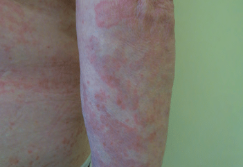
Urticarial vasculitis is a systemic disease characterized by the appearance of hives on the skin that persist for more than 24 hours and histologic signs of leukocytoclastic vasculitis. ICD-10 Code: L95.9.
The prevalence of the disease is 5 cases per million per year. Women between the ages of 30 and 50 are affected in 60-80% of cases, men less frequently, and children very rarely. The disease is observed in 5% of patients with chronic urticaria.
It is believed that urticarial vasculitis, similar to allergic vasculitides of the skin, is caused by the formation and deposition of immune complexes in the vessel, activation of the complement system, and degranulation of mast cells due to the action of anaphylatoxins C3a and C5a. Immune complex allergic reactions occur 1-2 weeks after contact with the antigen.
It is currently unknown whether the disease is hereditary, but one case has been reported in monozygotic twins, and recent studies of two cases of autosomal recessive hypocomplementemic vasculitis have identified homozygous mutations in DNAse1L3, which encodes an endonuclease. In most cases, urticarial vasculitis is idiopathic, but in some cases it has been associated with medication use, infections, environmental factors, and certain medical conditions.
Disease Triggers:
- Infections (Hepatitis B and C, Infectious Mononucleosis, and Lyme Disease)
- Medications (Cimetidine, Diltiazem, Procarbazine, Potassium Iodide, Fluoxetine, Procainamide, Cimetidine, Etanercept, Infliximab, Cocaine, Methotrexate, and Nonsteroidal Anti-Inflammatory Drugs)
- Serum Sickness
- Physical Factors (Physical exertion, exposure to heat or cold)
Associated Conditions:
- Autoimmune Diseases (Systemic Lupus Erythematosus, Sjögren's Syndrome)
- Hematologic Disorders (Plasma Cell Dyscrasias (IgM, IgG, IgA), Castleman's Disease, Cryoglobulinemia, and Idiopathic Thrombocytopenia)
- Cancer (Hodgkin Lymphoma, Acute Non-Lymphocytic Leukemia, Acute Myeloid Leukemia, Multiple Myeloma (IgA), Adenocarcinoma of the Colon, Kidney Cancer)
The disease is characterized by the eruption of multiple, well-defined wheals, often round in shape, sometimes with a fairly firm consistency, that persist in the same location for more than 24-48 hours (usually 3-5 days, sometimes up to 3 weeks). During this period, the location, shape and size of the rash elements hardly change. The color of the eruptions is red at first, gradually turning greenish-yellow as the process resolves, and they blanch with diascopy. After the wheals disappear, a hemorrhagic rash resembling "cayenne pepper" remains, which does not change with diascopy.
The rash is primarily localized on the trunk and proximal parts of the limbs, less commonly on the face. The rash elements usually coalesce to form circular, arcuate, or intricately shaped plaques. In 30-40% of cases, the eruptions are associated with angioedema. Erythematous macules, Raynaud's phenomenon, and blistering eruptions are occasionally observed. Subjectively, patients may experience burning, pain, tightness and, less commonly, itching in the affected areas.
Systemic Involvement in Urticarial Vasculitis
Common Symptoms:
- Fever (10-15%), malaise, and myalgia.
- Joints (60-70%): Arthralgia, polyarthritis (involving knee, ankle, elbow, wrist, metacarpophalangeal, and interphalangeal joints), synovitis.
- Abdominal Symptoms (20-30%): Nausea, vomiting, abdominal pain, gastrointestinal bleeding, or diarrhea. Splenomegaly or hepatomegaly.
- Eyes (over 10%): Episcleritis, uveitis, scleritis, conjunctivitis. Lungs (5-20%): Cough, dyspnea, or hemoptysis.
- Kidneys (10-20%): Glomerulonephritis, transient or persistent microscopic hematuria and proteinuria.
- Lymphadenitis (5%)
- Central Nervous System (rarely): Intracranial hypertension, optic nerve atrophy.
- Urticaria for at least 6 months.
- Hypocomplementemia.
- Histologically proven vasculitis.
- Arthralgia or arthritis.
- Uveitis or episcleritis.
- Glomerulonephritis.
- Intermittent abdominal pain.
- Reduced C1q or presence of anti-C1q autoantibodies.
- Complete blood count: Frequently elevated ESR.
- Urinalysis: Microhematuria and proteinuria in 10% of cases.
- Blood biochemistry: AST, ALT, bilirubin, creatinine, blood urea nitrogen.
- Testing for antinuclear antibodies, cryoglobulins, circulating immune complexes.
- Blood tests for rheumatoid factor and CRP (C-reactive protein).
- The levels of C1q, C3, C4 and CH50 are measured 2 or 3 times over several months.
- EIA for hepatitis B and C viruses, and Lyme disease.
- Chest X-ray.
- Consultation with an ophthalmologist.
- Urticaria
- Insect bites and papular urticaria.
- Hereditary and acquired angioedema.
- Serum sickness-like reaction.
- Erythema annulare centrifugum
- Erythema gyratum repens
- Erythema marginatum
- Adult-onset Still's disease.
- Schnitzler syndrome.
- Muckle-Wells syndrome (cryopyrin-associated periodic syndrome).
- Sweet syndrome.
- Kawasaki disease.
- Viral exanthems.
- Capillaritis.
- Urticarial phase of bullous pemphigoid.
- Atypical erythema multiforme.
- Rheumatoid neutrophilic dermatitis.
First-Line Therapy
- Antihistamines:
- Desloratadine 5 mg per day.
- Levocetirizine 5 mg per day.
- Loratadine 10 mg per day.
- Fexofenadine 120-180 mg per day.
- Cetirizine 10 mg per day.
- Ebastine 10-20 mg per day.
- Rupatadine 10 mg per day.
- Ranitidine 150 mg orally twice a day.
- Doxepin 10-25 mg 2-3 times a day.
- Nonsteroidal Anti-Inflammatory Drugs (NSAIDs):
- Indomethacin at 75-200 mg/day orally.
- Ibuprofen at 1600-2400 mg/day orally.
- Naproxen at 500-1000 mg/day orally.
Second-Line Therapy
- Colchicine 0.6 mg orally 2-3 times a day.
- Dapsone 50-150 mg/day orally.
Third-Line Therapy
- Prednisone 20-60 mg per day.
- Intravenous human immunoglobulin.
Fourth-Line Therapy
- Immunosuppressants: azathioprine, cyclophosphamide, cyclosporine, mycophenolate mofetil, methotrexate.
- Biological therapy: anakinra, anakinumab, tocilizumab, omalizumab, rituximab.

