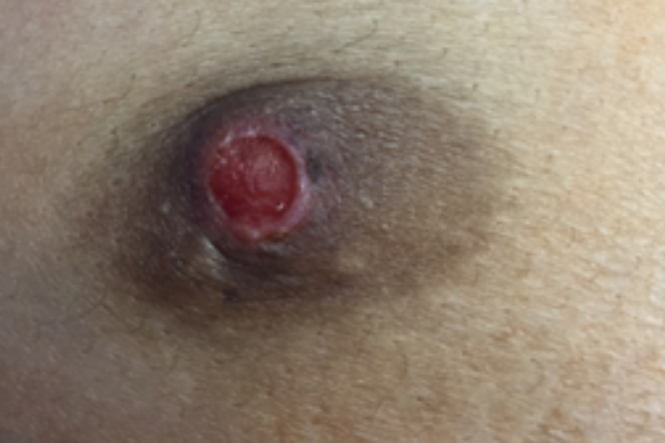
Nipple adenoma (also known as florid papillomatosis nipple, papillary adenoma nipple, erosive adenomatosis nipple, or blooming papillomatosis nipple) is a benign epithelial tumor that affects the ducts of the breast nipple. It is classified under the ICD-10 code D24.
The disease occurs between the ages of 20 and 50 (with an average age of 40-45), predominantly in women, less frequently in men and adolescents. The etiology is unknown. It is thought to be the result of uncontrolled hormonal hyperplasia. It is considered a tubular apocrine adenoma with a benign proliferative process in the milk ducts of the nipple.It is characterized by the appearance of a single, moderately elastic, red-colored papule with a smooth surface in the area of the nipple or areola of the breast. As the process progresses, the papule increases in size to a small nodule, becomes dense with a papillomatous or eroded surface, often covered with gray-yellow crusts. Peripheral erythema is usually observed around the element. Serous or bloody discharge from the nipple, which increases during the premenstrual period, is characteristic.
In severe cases, the entire nipple can become swollen, hardened, enlarged, and deformed. Unilateral formation is almost always observed, bilateral lesions are described in some cases. Pain and itching are commonly reported. The disease is chronic and does not tend to resolve spontaneously.The diagnosis is made based on the clinical picture and histological examination. The histological picture is quite diverse. Dermoscopy reveals linear, rarely serpiginous or ring-shaped cherry-red structures, which are believed to represent altered ductal openings of the nipple.
Histological findings:- Nodular florid epithelial proliferation of the lactiferous ducts of the nipple with papillary hyperplasia / adenosis / sclerosis / mixed pattern
- Epithelium is cytologically bland
- Myoepithelial cell layer is preserved uniformly
- 4 major morphologic patterns:
- Sclerosing papillary hyperplasia pattern:
- Intraductal papillary growth with prominent stromal proliferation (collagenous bands, myxoid change or elastosis)
- Focal central necrosis may be present
- Squamous cysts can be present in duct orifices
- Papillary hyperplasia pattern:
- Papillary growth in large ducts with less prominent stromal proliferation and focal necrosis
- Replacement of epidermis with glandular epithelium
- Adenosis pattern:
- Small duct proliferation with a pattern resembling sclerosing adenosis and may be pseudoinfiltrative
- Necrosis is uncommon
- Mixed pattern:
- Any combination of the above described patterns
- Sclerosing papillary hyperplasia pattern:
- May be in continuity with the skin surface, which may appear eroded or ulcerated
- Fairly well circumscribed but unencapsulated
- Other features often seen:
- Squamous metaplasia and superficial keratin cysts
- Acanthosis
- Toker cell hyperplasia in the epidermis
- Multinucleated giant cells
- Apocrine metaplasia
- In rare cases, ductal carcinoma in situ involving an adenoma or a contiguous invasive carcinoma have been reported
Cited: Sabljic T, Mrkonjic M. Nipple adenoma. PathologyOutlines.com website. www.pathologyoutlines.com/topic/breastnipplead.html. Accessed January 13th, 2024.
- Paget's disease
- Nipple eczema
- Acrochordon
- Hyperkeratosis of the nipple
- Glands of Montgomery (Areolar glands)
- Syringocystadenoma papilliferum

