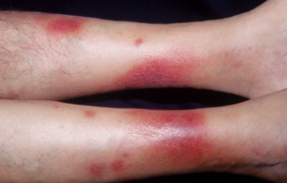
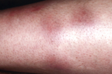
Erythema nodosum is an acquired acute or chronic condition characterized by the presence of deep, erythematous, painful nodules typically located on the extensor surfaces of the lower limbs. ICD-10 Code: L52.
Erythema nodosum affects individuals of all races and both sexes and is not genetically inherited. It is considered to be an idiopathic disorder and the hypothesis that it is a cutaneous manifestation of an immune response still requires clarification. Erythema nodosum can be seen as a reaction arising from a variety of causes. Immune mechanisms are thought to mediate this reaction, which develops in response to various antigenic stimuli, including viral, bacterial (such as streptococcus), mycobacterial, deep fungal infections (coccidioidomycosis and blastomycosis), sarcoidosis, lymphoma, inflammatory bowel disease, and medications (especially oral contraceptives and possibly halogens).
Often, Yersinia infection is the underlying disease. The typical pathogenetic mechanism in this case involves the host immune response to circulating immune bodies, which can lead to the formation of immune complexes that contribute to the disease. In inflammatory bowel disease, erythema nodosum results from increased intestinal permeability to endo- and exogenous antigens.
Erythema nodosum can also be induced by radiotherapy. Products of the degradation of irradiated cancer cells can activate circulating antibodies and complement, leading to the formation of immune complexes and thus triggering the response. Radiation therapy is known to exacerbate certain reactive dermatoses.Acute Erythema Nodosum
Chronic Erythema Nodosum
Chronic erythema nodosum is characterized by a persistent recurrent course and usually occurs in mature women. Exacerbations are more common in spring and autumn and are characterized by the appearance of a small number of bluish-pink, dense, subcutaneous, slightly painful nodules the size of a walnut or hazelnut, which merge into plaques with resolution in the center and persist for several months. In the early stages, the nodules may not change the color of the skin, may not protrude above the skin, and may only be palpable. Almost exclusively, the nodules are located on the shins (usually their anterior and lateral surfaces). There is mild swelling of the shins and feet. General symptoms are inconsistent and mild. Episodes may last several months, during which time some nodules may disappear while others appear in their place.
Erythema nodosum migrans (Vilanova Disease)
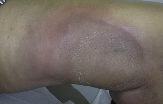
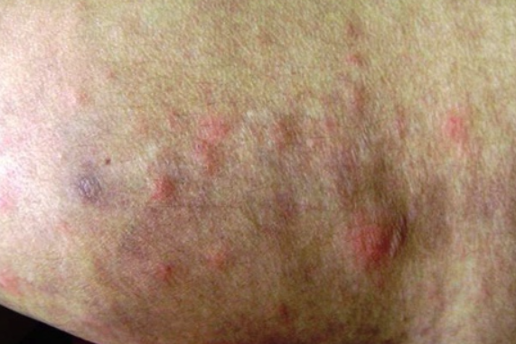
A chronic variant of the disease has been described, called "subacute migratory panniculitis" ("Vilanova disease"). In this case, one or more nodules appear, typically unilateral. The consistency of these nodules is less dense than in chronic erythema nodosum, and they tend to persist for longer. These nodules expand slowly at the periphery and as they dissolve in the center, they assume a ring-like shape. The skin at the periphery of the nodule has a bright red color, while the center appears bluish brown. The diameter of these annular elements can reach 10 cm or more.
There is also a superficial infiltrative form of erythema nodosum migrans characterized by flat superficial sclerodermoid-like infiltrates that tend to enlarge peripherally.Erythema nodosum leprosum
- Erythema induratum (nodular vasculitis)
- Superficial migratory thrombophlebitis
- Cutaneous polyarteritis nodosa
- Sweet's syndrome
General therapeutic recommendations
- Bed rest is recommended, which may lead to spontaneous resolution of the condition within a relatively short period of time. Since the condition often resolves spontaneously, the most important aspect is to identify and treat the underlying cause.
- In general, erythema nodosum is a relatively common and usually benign condition. It is usually diagnosed clinically. Treatment is primarily symptomatic and includes bed rest, nonsteroidal anti-inflammatory drugs, and potassium iodide. As the disease tends to resolve spontaneously, the main challenge is to identify and treat the underlying cause of the condition.
Treatment of the Acute Form
Potassium iodide (a preparation consisting of 76% iodine and 23% potassium) is the treatment of choice. After oral ingestion, potassium iodide is rapidly absorbed in the gastrointestinal tract and distributed in the intercellular space. Iodine is concentrated in the thyroid and other tissues such as salivary glands, gastric mucosa, and mammary glands. Potassium iodide can act relatively quickly, making it easy to assess the effectiveness of treatment. The drug is administered orally at a dose of 300 mg three times a day. Complete resolution of lesions occurs within 10-14 days of starting the drug, especially if treatment is started promptly after the onset of the disease. The best response is observed in patients with erythema nodosum associated with systemic symptoms such as fever, arthralgia, and the presence of C-reactive protein. Nausea and vomiting may occur occasionally, but usually does not require discontinuation of treatment. Adverse skin reactions may include erythematous, purpuric, urticarial, acneiform, nodular, pustular, and vegetative lesions. In addition to these relatively minor side effects, more serious adverse reactions may occur in pregnant women and in patients with a history of kidney or thyroid disease. Severe hypothyroidism secondary to exogenous iodide intake has been reported, resulting from inhibition of its organic binding in the thyroid gland due to an excess of iodide, a phenomenon known as the Wolff-Chaikoff effect.
The mechanism by which potassium iodide works is not fully understood. One hypothesis is that potassium iodide may trigger the release of heparin from fat cells, and heparin, in turn, suppresses cellular immunity. The drug may also modulate neutrophil function by inhibiting the production of hydrogen peroxide and hydroxyl radicals. These oxygen intermediates are so reactive that they can damage tissue. By preventing the production of oxygen intermediates, potassium iodide protects tissues from self-oxidative damage.
In cases of acute form with fever and joint pain, treatment with colchicine at a dose of 1 mg/day for one month is carried out.
Erythema nodosum may indicate the presence of sarcoidosis. In 80% of patients with sarcoidosis, the disease resolves spontaneously and treatment is rarely required. Treatment is the same as for the acute asymptomatic form, as described above.
Erythema nodosum as a symptom of gastrointestinal infections is treated with erythromycin at a dose of 250 mg four times a day for 4-6 weeks.
Erythema nodosum caused indirectly by oral contraceptives often resolves when the contraceptive is discontinued.
Acute forms of hepatitis B and C are sometimes associated with erythema nodosum. The nodules typically resolve slowly after the acute phase of the disease. There have been reports of a strong association between hepatitis B vaccination and the onset of the condition, with spontaneous resolution of the lesions.
Treatment of the chronic form
Erythema nodosum is a debilitating chronic disease for which many treatment options have been proposed, but none are generally effective. Although patients respond rapidly to corticosteroids, the risk of their chronic use necessitates the search for an alternative approach.
In chronic cases, high doses of aspirin often fail to provide a response, but they can respond significantly to the anti-inflammatory effect of indomethacin at doses of 100-150 mg/day. The suppression of the process by indomethacin is related to blocking the synthesis of prostaglandins in subcutaneous adipose tissue, which affects both cellular and humoral immune responses.
Chronic forms can also be successfully treated with naproxen. However, indomethacin outperforms systemic corticosteroids and may have advantages over other less potent nonsteroidal anti-inflammatory drugs.
Hydroxychloroquine is an effective and safe alternative therapy for some patients at a dose of 200 mg twice daily, then tapered to 200 mg once daily for 4 months.
Treatment of erythema nodosum leprosum
Currently, thalidomide is recommended as first-line therapy for erythema nodosum leprosum . Sheskin analyzed data from 4522 cases of leprosy erythema nodosum and found the drug to be 99% effective, even though thalidomide has no direct effect on Mycobacterium leprae. Clearance occurs within 24-48 hours after initiation of thalidomide therapy. In addition, the drug has been reported to have a beneficial effect on reactive polyneuritis. Erythema nodosum leprosum is associated with elevated levels of TNF-alpha and interferon-gamma. After thalidomide therapy, the serum level of TNF-alpha decreased, as did the number of helper T cells. Thalidomide is known for its inhibitory effect on TNF-alpha. The optimal dose for a rapid response is 400 mg/day, followed by maintenance therapy at a dose of 100 mg/day. The duration of treatment varies from several weeks to several years. There have also been reports of the efficacy of plasma exchange and the absence of relapses. This method has been used when traditional treatments have been ineffective.


