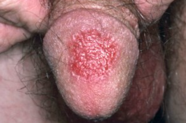
Fixed drug eruption (FDE) is a type of drug eruption characterized by recurrent eruptions on the same areas of the skin after taking certain medications. ICD-10 Code: L27.1.
This condition affects both sexes and all age groups, but is more common in individuals with dark or black skin. Fixed drug eruptions account for 14-22% of drug eruptions. The pathogenesis is not well understood. It is believed that the ingested drug activates CD8+ T lymphocytes, which migrate into intraepidermal spaces and release gamma interferon and cytotoxins such as granzymes and perforins. This leads to an increase in neutrophils, CD4+ T cells, mast cells and sometimes eosinophils at the site of the lesions, initiating apoptosis of basal keratinocytes. After discontinuation of the drug, activation of T lymphocytes stops. However, basal keratinocytes continue to produce IL-15, which allows some CD8+ T cells to recognize the antigenic structure of the drug. These cells reside in the upper layers of the dermis at the site of the previous lesion and become activated when the drug or a cross-reacting drug re-enters the body, resulting in the appearance of new eruptions in that area. Some authors believe that repeated lesions may occur under the influence of ultraviolet radiation. To date, more than 100 drugs and substances have been described that can cause a fixed drug eruption (FDE). In the vast majority of cases, the reaction occurs when these substances are taken orally, while eruptions are much less common with parenteral administration and topical application. The most commonly implicated agents in causing fixed drug eruption include:
- Sulphonamides (sulfamethoxazole/trimethoprim) and drugs containing sulphonamide moieties (oral hypoglycemic agents and diuretics) - accounting for up to 75% of cases.
- Antibiotics (tetracycline, minocycline, amoxicillin, vancomycin, teicoplanin, fluoroquinolones).
- Non-steroidal anti-inflammatory and analgesic drugs (acetylsalicylic acid, sodium metamizole, phenylbutazone, paracetamol).
- Sedative medications (barbiturates, glutethimide).
- Anticonvulsants (phenytoin).
- Antiparasitic and antifungal drugs (metronidazole, ornidazole, tinidazole, nystatin, fluconazole).
- Quinine and quinidine.
- Oral contraceptives.
Classic fixed drug eruption
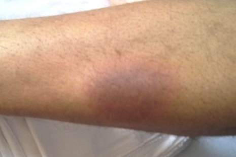
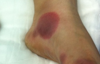
After initial use of the medication, typically within 1-2 weeks, a bright red, circular or oval-shaped spot with clear borders and a diameter of 0.5 to 10 cm appears. The development of the spot is often accompanied by itching or burning. Within 24 hours, there is swelling and the formation of a slightly raised plaque on the surface of the skin that changes color over several days to dark red, brownish-blue, or dark purple. Approximately 2-3 weeks after discontinuing the medication, the inflammatory symptoms subside and the plaque spontaneously resolves without a trace or with post-inflammatory hyperpigmentation in the form of a brown-purple spot that fades and disappears over several weeks to months. Eruptions may occur anywhere on the body and on mucous membranes.
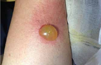
With readministration of the same drug or a cross-reacting drug, new patches (while previous ones may increase in size) appear at previous eruption sites within 30 minutes to 24 hours (usually within an average of 8 hours).Bullous fixed drug eruption
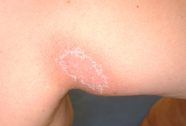
Non-pigmented fixed drug eruption
Genital fixed drug eruption
Diagnosis is based on the clinical picture and medication history.
Oral provocation test: Oral administration of the "suspected" drug and observation for the appearance of new eruptions or worsening of existing rash elements within 24 hours (prohibited in generalized bullous form).
Patch test: Application of the drug to the skin in areas where eruptions typically occur; this results in an inflammatory reaction in approximately 30% of patients.
- Erythema multiforme.
- Erythema dyschromicum perstans
- Large plaque parapsoriasis
- Syphilitic roseola.
- Pityriasis rosea.
- Pemphigus vulgaris
- Bullous pemphigoid
- Linear IgA dermatosis.
- Herpes simplex.
- Stevens-Johnson syndrome.
- Behçet's disease.
- Insect bites with hyperpigmentation.
- Post-inflammatory hyperpigmentation.
- Contact dermatitis.
- Erythema migrans (Lyme disease).
- Tinea corporis.
- Pigmented lichen planus
- Diabetic bulla.
Identification and discontinuation of the etiologic agent is required. Afterward, the rash spontaneously resolves within 2-3 weeks. Topical steroids and oral antihistamines may be used for symptomatic treatment. There are no effective treatments for residual hyperpigmentation (hydroquinone is ineffective).
In cases of secondary infection of eroded elements, local or systemic antibiotic therapy may be required. Patients with the generalized bullous form may require hospitalization.
