Atopic Eczema (Atopic Dermatitis - AD) is a multifactorial inflammatory skin condition characterized by itching, a chronic relapsing course, and age-specific features of lesion localization and morphology. ICD-10 Code: L20
Genetics play an important role in the pathogenesis of atopic dermatitis, leading to skin barrier dysfunction, immune system defects (stimulation of T cells with subsequent overproduction of IgE), hypersensitivity to allergens and non-specific irritants, colonization by pathogenic microorganisms (Staphylococcus aureus, Malassezia furfur), and an imbalance in the autonomic nervous system with an increase in inflammatory mediators.
Atopic dermatitis is one of the most common skin diseases, accounting for 20% to 40% of all skin diseases, occurring in all countries, in individuals of both sexes and in different age groups. The incidence has increased 2.1-fold in the last 16 years. The prevalence of the disease in the pediatric population is up to 20%, and in adults it ranges from 1% to 3%.
Atopic dermatitis develops in 80% of children if both parents have the disease and in over 50% of children if only one parent has the disease, with the risk of developing the disease increasing one and a half times if the mother has the disease.
Early-onset atopic dermatitis (between 2 and 6 months of age) is observed in 45% of patients and within the first year of life in 60% of patients. Spontaneous remission occurs in 65% of children with atopic dermatitis by the age of 7 years, and in 74% of children with atopic dermatitis by the age of 16 years. Between 20% and 43% of children with atopic dermatitis go on to develop bronchial asthma, and twice as many develop allergic rhinitis.Age-specific features of localization and morphology of skin elements distinguish atopic dermatitis from other eczematous and lichenoid skin diseases. The main differences in clinical manifestations between age groups are the localization of affected areas and the ratio of exudative and lichenoid components. Pruritus is a constant symptom of the disease in all age groups.
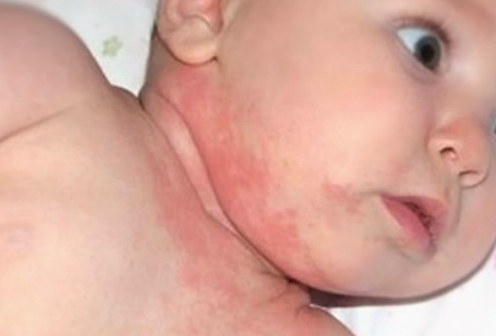
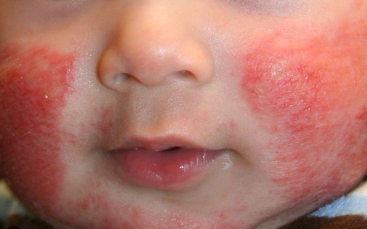
Infancy
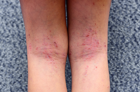
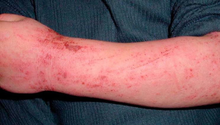
Childhood
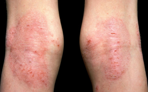
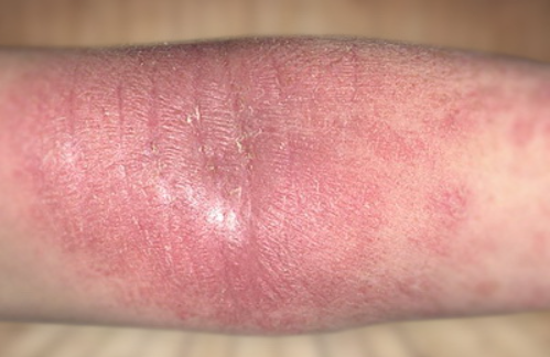
Adult
Clinically, atopic dermatitis can be divided into:
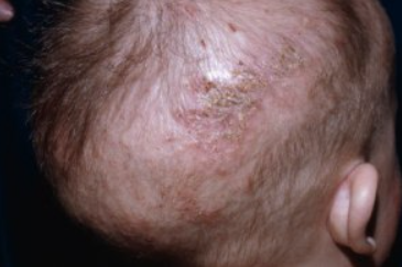
Exudative ("wet") atopic eczema
Erythematous-squamous atopic eczema
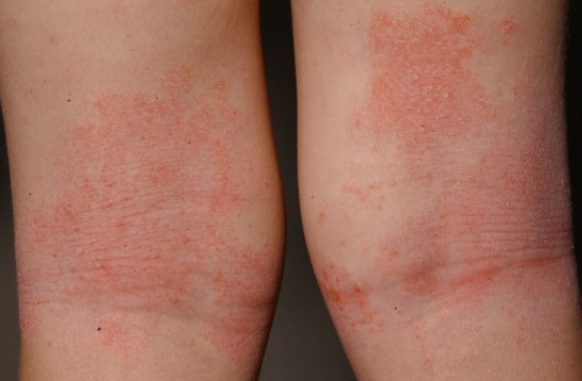
Erythematous-squamous form is more common in children aged 1.5 to 3 years and is characterized by pruritic papules, erosions, and excoriations, as well as mild erythema and infiltration in the areas of eruption on the skin of the trunk, upper and lower limbs, and less frequently on the face. Dermographism is pink or mixed.
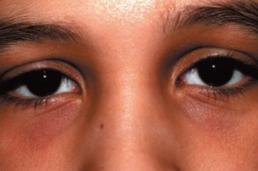
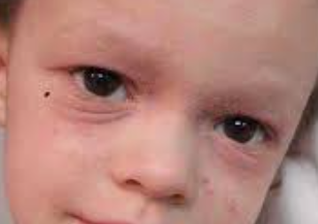
Erythematous-squamous form with lichenification is observed in children as young as 3 years and in adults. It is characterized by erythematous-squamous and papular lesions. The skin is dry, lichenified, with numerous excoriations and fine, plaque-like scales. Eruptions are most common on the flexural surfaces of the limbs, the dorsal surface of the hands, and the anterior and lateral aspects of the neck. Hyperpigmentation of the periorbital skin may occur, along with the appearance of a fold under the lower eyelid (Dennie-Morgan lines). Increased dryness of the skin is noted. Dermographism is white and persistent or mixed. Pruritus is marked, constant, occasionally paroxysmal.
Lichenoid atopic eczema
Pruritic atopic eczema
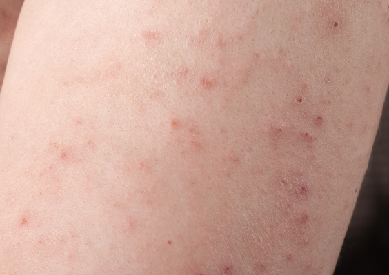
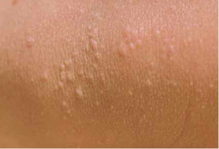
Papular form of atopic eczema
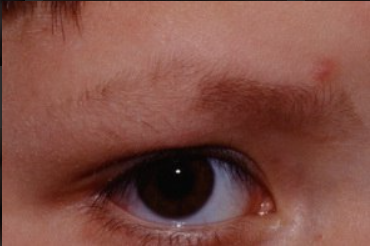
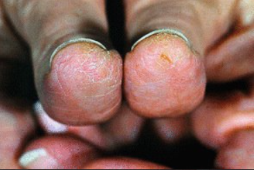
Clinical Symptoms of Atopic Eczema
Diagnosis of atopic dermatitis is established based on the patient's medical history and the characteristic clinical presentation.
Diagnostic Criteria (The Hanifin-Rajka criteria)
Main diagnostic criteria:
- Skin itching.
- Skin involvement: in infants, rash on the face and flexor surfaces of the limbs; in children and adults, lichenification and scratch marks in the folds of the extremities.
- Chronic relapsing course.
- Presence of atopic diseases in the patient or their relatives.
- Onset of the disease in early childhood (before 2 years).
Additional diagnostic criteria:
- Seasonal exacerbations (worsening in cold weather and improvement in summer).
- Exacerbation triggered by provoking factors (allergens, irritants, food, emotional stress, etc.).
- Elevated levels of total and specific IgE in the blood serum; peripheral blood eosinophilia.
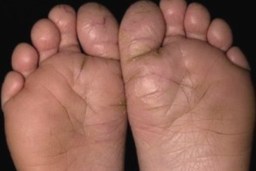
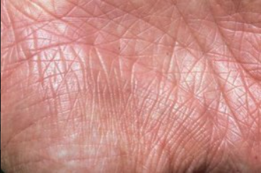
- Hyperlinear palms and soles.
- Follicular hyperkeratosis
- Pruritus with increased sweating.
- Dry skin (xerosis).
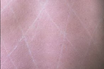
- White dermographism.
- Tendency toward cutaneous infections.
- Tendency towerd hand and foot dermatitis
- Nipple eczema.
- Recurrent conjunctivitis.
- Hyperpigmentation of the skin in the periorbital area.
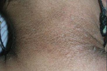
- Folds on the front of the neck.
- Dennie-Morgan sign
- Cheilitis.
To diagnose atopic dermatitis, a combination of three main criteria and at least three additional criteria is required.
Mandatory laboratory tests include:
- Complete blood count.
- Urinalysis.
- Blood Biochemistry.
Additional laboratory tests include:
- Determination of total IgE levels in serum
- Allergological testing of blood serum - determination of specific IgE to food, household antigens, antigens of plant, animal etc.
- Seborrheic dermatitis
- Allergic contact dermatitis
- Diaper dermatitis
- Psoriasis
- Ichthyosis vulgaris
- Nummular eczema
- Tinea corporis
- Mycosis fungoides (early stages)
- Lichen simplex chronicus
- Actinic reticuloid
- Phenylketonuria
- Enteropathic acrodermatitis
- Wiskott-Aldrich syndrome.
Treatment Objectives
- Achieving clinical remission of the condition.
- Eliminating or reducing inflammation and skin itching, preventing and treating secondary infections, moisturizing and softening the skin, restoring its protective properties.
- Improving the quality of life for patients.
An important aspect of treatment is the elimination of triggering factors (psychological stress, dust mites, molds, climatic changes, environmental pollution, dietary disorders, improper skin care routines, irrational use of synthetic detergents as well as shampoos, soaps and lotions with high pH values, tobacco smoke, etc.). The patient's medical history, clinical manifestations of the disease and examination results are analyzed to evaluate the significance of specific factors for the individual patient and to take measures to eliminate them.
All patients with atopic dermatitis, regardless of the severity, extent, acuteness of the skin process or the presence of complications, are prescribed basic skin care products.
For localized skin involvement, mild to moderate severity, and during exacerbations, topical therapy is primarily used. This may include potent or moderately potent topical corticosteroids and/or topical calcineurin inhibitors in addition to basic therapy.
After resolution of the exacerbation, topical corticosteroids and calcineurin inhibitors are discontinued and the patient continues to receive only basic therapy.
For moderate atopic dermatitis, phototherapy and, if indicated, detoxifying agents may be prescribed during exacerbations.
Treatment of patients with severe atopic dermatitis may include systemic medical therapy or phototherapy in addition to topical treatments.
The effectiveness of topical therapy depends on three basic principles: sufficient drug potency, adequate dosage, and proper application. Topical medications should be applied to moist skin. Topical anti-inflammatory medications are applied directly to the affected areas of skin and discontinued when the condition is resolved. Recently, proactive treatment has been recommended: prolonged use of low-dose topical anti-inflammatory agents on affected skin areas, combined with the use of emollients on the entire skin surface, and regular visits to the dermatologist for evaluation of the skin condition.
The amount of topical medication for external use is measured using the Finger Tip Unit (FTU) rule. One FTU is equal to a column of ointment 5 mm in diameter and equal in length to the distal phalanx of the index finger, weighing approximately 0.5 grams. This dose of topical agent is sufficient to apply to the skin of two adult palms, covering approximately 2% of the total body surface area.
Topical Corticosteroids
Topical corticosteroids (TCS) are the first-line agents for local anti-inflammatory therapy and have a significant effect on the skin condition compared to a placebo effect, especially when used with wet-dry dressings. Proactive TCS therapy (use twice a week for 3-4 months) helps to reduce the likelihood of exacerbations. TCS may be recommended in the early stages of AD exacerbations to reduce itching.
The use of TCS is indicated for severe inflammatory manifestations, significant pruritus and when other topical therapies have failed. TCS should be applied only to the affected areas of skin, leaving healthy skin untouched. TCS are classified based on the composition of active ingredients (single and combined) and the strength of their anti-inflammatory activity.
- Fluticasone propionate, cream, 0.05%, applied thinly to affected skin areas 2 times a day for 2 weeks.
- Triamcinolone acetonide, ointment, 0.025%, 0.1%, applied thinly to affected skin areas 2 times a day for 2 weeks
- Clobetasol propionate, ointment, 0.05%, applied thinly to affected skin areas 2 times a day for 2 weeks.
- Betamethasone valerate, cream, ointment, applied 2 times a day thinly to affected skin areas for 2 weeks.
- Mometasone furoate, cream, ointment, 0.1%, applied thinly to affected skin areas 2 times a day for 2 weeks.
Topical Calcineurin Inhibitors
Topical calcineurin inhibitors are an alternative to topical corticosteroids and the preferred treatment for atopic dermatitis on sensitive areas of the body (face, neck, folds). These preparations are also recommended when a patient does not respond to topical corticosteroid therapy.
Pimecrolimus is used for the topical treatment of mild to moderate atopic dermatitis on affected areas, for short or long periods, in adults, adolescents, and children over 3 months of age.
- Tacrolimus, cream, ointment, 0.1%, applied thinly to affected skin areas once a day for 2-4 weeks.
- Pimecrolimus, cream, ointment, 1%, applied thinly to affected skin areas once a day for 2-4 weeks.
Ultraviolet therapy:
- Narrowband ultraviolet at 311 nm (UVB range, wavelength 310-315 nm with an emission peak at 311 nm).
- UVA-1 range, wavelength 340-400 nm.
- Broadband ultraviolet therapy in the UVB range with a wavelength of 280-320 nm.
Systemic therapy:
Cyclosporine
- Cyclosporine 3-5 mg per kg per day in two divided doses with a 12-hour interval.
- During cyclosporine treatment, it is important to systematically monitor kidney and liver function, blood pressure, plasma levels of potassium and magnesium (especially in patients with kidney impairment), as well as urea, creatinine, uric acid, bilirubin, liver enzymes, amylase, and lipid levels in the blood serum.
Systemic glucocorticoids
Systemic glucocorticoids are used in the treatment of patients with atopic dermatitis only for the management of acute exacerbations in severe cases of the disease in adults and extremely rarely in children. This prescribing strategy is primarily associated with the possibility of disease exacerbation after discontinuation of the medication. Prolonged use of systemic glucocorticoid medications also increases the likelihood of developing side effects.
For the treatment of acute exacerbations, intravenous prednisone is administered according to the following schedule Day 1/the first two days: 90 mg in the morning, followed by 60 mg in the morning for the next two days, and then, if necessary, prednisone may be administered at a dose of 30 mg for another 2-3 days with subsequent discontinuation.
Antihistamine Drugs
- Loratadine 10 mg orally once a day for 10-20 days or
- Cetirizine 10 mg orally once a day for 10-20 days or
- Levocetirizine 5 mg orally once a day for 10-20 days or
- Desloratadine 5 mg orally once a day for 10-20 days or
- Clemastine 1.0 mg orally twice a day or 2.0 mg intravenously or intramuscularly twice a day for 10-20 days or
- Mebhydroline 100 mg orally twice a day for 10-20 days or
- Fexofenadine 120-180 mg orally once a day for 10-20 days.
Basic therapy includes the regular use of moisturizers and emollients and the elimination (if possible) of triggering factors.
Educational programs
Educational programs are highly effective and are used in many countries as part of schools for people with atopic dermatitis.
Moisturizers/Emollients
General recommendations for the use of moisturizers and emollients are as follows:
- Patients with atopic dermatitis should use moisturizers and emollients continuously, frequently and in large amounts (at least 3-4 times per day), both independently and after water treatments using the "soak and seal" method: daily baths with warm water (27-30°C) for 5 minutes with the addition of bath oil (2 minutes before the end of the water treatment), followed by the application of an emollient to moist skin (after water treatments, gently pat the skin dry, avoiding rubbing). However, there is evidence that applying emollients without a bath has a longer lasting effect.
- The most pronounced effect of moisturizers and emollients is observed when they are used continuously in the form of creams, ointments, bath oils and soap substitutes. In winter it is better to use richer ingredients. To achieve a clinical effect, it is necessary to use a sufficient amount of emollients (an adult with extensive skin involvement can use up to 600 grams per week, while a child can use up to 250 grams per week).
- Emollient creams should be applied either 15 minutes before or 15 minutes after using an anti-inflammatory, especially if the emollient has a greasier base.
- Consistent use of moisturizers and emollients can help eliminate dryness, itching and inflammation, thereby limiting the use of topical glucocorticoids and achieving short- and long-term steroid-sparing effects (reducing TGC dosage and the likelihood of side effects) in mild and moderate cases of atopic dermatitis (AD).
- Emollients may be applied immediately after application of the topical calcineurin inhibitor pimecrolimus. However, moisturizers and emollients should not be applied for 2 hours after the application of topical tacrolimus. After water procedures, emollients should be applied before applying calcineurin inhibitors.
- Side effects from the use of emollients are rare, but there have been cases of contact dermatitis and occlusive folliculitis. Some lotions and creams may be irritating due to the presence of preservatives, solvents, and fragrances in their composition. Water-based lotions may cause dryness due to evaporation.
Topical Antibiotics
Topical antibacterial agents are used to treat localized forms of secondary infections. Topical combination products containing glucocorticoids in combination with antibacterial, antiseptic and antifungal agents may be used for short courses (usually within 1 week) when signs of secondary skin infection are present.
Topical antibiotics should be applied to the affected skin areas 1-4 times daily for up to 2 weeks, depending on the clinical manifestations.
Antiviral Drugs
One of the serious and life-threatening complications of atopic dermatitis is the development of eczema herpeticum when the skin is infected with herpes simplex virus type I. This condition requires systemic antiviral therapy with acyclovir or other antiviral drugs.
The recommended systemic antiviral drugs for the treatment of eczema herpeticum in children are acyclovir,valacyclovir or famcyclovir. In case of disseminated processes accompanied by general symptoms (elevated body temperature, severe intoxication), hospitalization is necessary. In a hospital, intravenous administration of acyclovir is recommended. Topical therapy includes the use of antiseptic agents.

