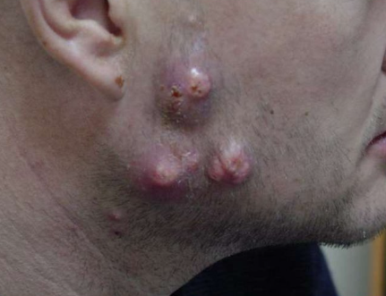
Bacillary angiomatosis is an opportunistic cutaneous or systemic infection in severely immunosuppressed individuals caused by the gram-negative bacteria Bartonella henselae and Bartonella quintana. It is characterized by the development of angiomatous papules, nodules, or plaques and involvement of internal organs. ICD-10 code is A79.8.
The disease is mainly observed in HIV-infected individuals, less frequently in patients with other immunodeficiencies, and rarely in immunocompetent individuals. The incidence is estimated at 1.2-1.42 cases per 1000 AIDS patients, with a higher incidence in men aged 40-60 years. Infection with B. henselae occurs through scratches and bites from cats, dogs, rabbits, and monkeys. Transmission by arthropods has not been ruled out. Infection with B. quintana is observed in homeless individuals and is associated with bites from head or body lice.
In immunosuppressed patients (CD4 cell count below 200 cells/mm³), B. henselae may cause disseminated "cat scratch disease" characterized by granulomatous lymphadenitis. Patients with bacillary angiomatosis caused by B. quintana present with subcutaneous nodules and involvement of the liver, spleen, and bones. Bacillary angiomatosis usually occurs after mild or asymptomatic bacteremia in HIV-infected individuals, organ transplant recipients, and patients receiving cytostatic therapy. It is rare in healthy individuals. The mechanism of disease development in HIV-infected individuals is not well understood.
The incubation period ranges from a few days to 2-3 months (average of 25-30 days).
Skin lesions present in various forms:
- Smooth, dome-shaped angiomatous papules of reddish, bluish, or brownish color.
- Exophytic reddish papules, often ulcerated in the center and covered with crusts, which may bleed easily.
- Subcutaneous nodules covered with normal skin.
- Bluish-red infiltrates of the skin.
- Dry, hyperkeratotic erythematous plaques over areas of osteoporosis.
Lesions of bacillary angiomatosis are rarely solitary; they are more often multiple and may occur on any part of the skin except the palms, soles, and oral mucosa. The number of eruptions may reach several hundred. On palpation, the rash elements are tender and have a soft or firm consistency. Subsequent hematogenous and lymphatic dissemination of Bartonella spp. may lead to involvement of soft tissues, internal organs, bone marrow, lymphadenopathy, hepatomegaly, and splenomegaly. Blood-filled cysts known as bacillary peliosis may develop in the liver and spleen.
In HIV-infected individuals, the following syndromes can occur:
- Bacillary angiomatosis of the skin
- Peliosis hepatis (involvement of the liver)
- Parenchymal peliosis (involvement of the liver and spleen)
- Bartonella sepsis (fever and bacteremia)
The course of bacillary angiomatosis is variable. In some patients, the lesions resolve spontaneously, but more commonly, death may occur from laryngeal obstruction, liver failure, or pneumonia if untreated. Like other opportunistic infections in HIV infection, bacillary angiomatosis is prone to recurrence.
Polymerase chain reaction (PCR) can detect Bartonella DNA in infected tissue.
Enzyme-linked immunosorbent assay (ELISA) can detect Ig antibodies to Bartonella spp.
Blood biochemistry shows elevated levels of gamma-glutamyltransferase (GGT) and alkaline phosphatase in peliosis hepatis.
Radiologic and ultrasonographic studies may reveal bone and visceral pathology.
- Kaposi sarcoma
- Pyogenic granuloma
- Epithelioid (histiocytoid) hemangioma
- South American bartonellosis (Peruvian wart)
- Sclerosing hemangioma
- Cherry angioma
- Angiokeratomas
- Disseminated deep mycoses
- Disseminated mycobacterial infections
Antibiotics:
- Erythromycin, 500 mg orally four times a day.
- Doxycycline, 100 mg orally twice a day.
- Ciprofloxacin, 750 mg orally twice a day.
- Azithromycin, 500 mg orally once a day.
Antibiotic treatment is continued until resolution of the rash, which typically ranges from 3-4 weeks to 2-4 months. Successful treatment with clarithromycin and rifampicin has been reported. Jarisch-Herxheimer reaction may occur shortly after initiation of treatment. Local therapy is indicated for solitary lesions: excision, electrocautery, or cryotherapy in combination with systemic therapy. Lifelong therapy is recommended for recurrent cases.

