Behçet's disease is a systemic vasculitis of unknown etiology. It is characterized by recurrent ulcerations of the oral cavity and genitalia, as well as involvement of the eyes, joints, gastrointestinal tract, central nervous system, and other organs. It has a chronic course with unpredictable exacerbations and remissions. ICD-10 code is M35.2.
Factors that can affect the development of the disease include:
- Gender: It more commonly affects men in the second or third decade of life.
- Ethnicity and geographical location: It is more prevalent in countries along the historical Silk Road.
Oral Ulcers (Aphthae)
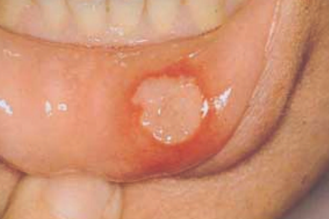
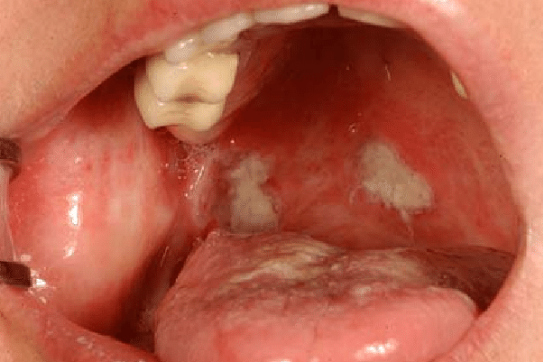
Oral ulcers are often the first and most common manifestation of Behçet's disease, with at least three recurrences per year. The ulcers may be solitary or multiple, painful, and have rounded or sharp erythematous borders that resemble punched-out lesions. The ulcers are covered with grayish white or yellowish fibrin (necrotic tissue). They are predominantly located in the anterior parts of the oral cavity, such as the buccal mucosa, lips, gums, and tongue. Posterior localization of aphthae is less common and involves the tonsils, soft and hard palate, and posterior pharyngeal wall.
Minor aphthae: These are more common, ranging from 1 to 5 in number and up to 1 cm in size. They are superficial, moderately painful, and heal without scarring within 4-14 days.
Large aphthae: These are less common and larger, exceeding 1 cm in size. They are deep, very painful, and can significantly interfere with the patient's daily activities. They heal in 2-6 weeks, often leaving scarring.
Herpetiform aphthae: These are the rarest type and appear as small, numerous ulcers that are 2-3 mm in size and painful.
Genital ulcers
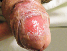
In men, they are located on the scrotum and penis; in women, they occur on the labia majora, labia minora, vulva, vagina, and cervix. Exacerbations often occur prior to menstruation.
The ulcers are similar to aphthae but occur less frequently, about 2-3 times a year. They are often painful but may be asymptomatic. In most cases, they leave white or pigmented scars. Perianal ulcers can occur in both men and women and are large and deep.
In men, epididymitis may occur simultaneously with genital ulcers, resulting in infertility. Due to the variety of clinical manifestations, patients may initially be seen by physicians of other specialties (non-rheumatologists), which often leads to a delayed diagnosis of Behçet's disease.
Skin lesions
Nodular erythema in women (present in more than 2/3 of patients) is localized on the anterior surface of the legs, ankles, face, hands, and buttocks. It appears as raised red nodules with subcutaneous induration. The nodules may ulcerate and resolve within 10-14 days without scarring, leaving hyperpigmentation after healing.
Other characteristic features include recurrent pseudofolliculitis, pustular and acne-like eruptions (more common on the back in men in the absence of glucocorticoid therapy), thrombophlebitis, bullous or necrotizing vasculitis, palpable purpura, and pyoderma gangrenosum.
Eye involvement
Young males are at high risk, while older females are at lower risk.
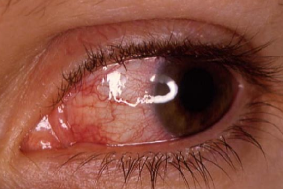
Typically, involvement is bilateral and recurrent episodes of inflammatory attacks are characteristic. Posterior uveitis has a poor prognosis for vision, with manifestations including periarteritis and periphlebitis with occlusive retinal vasculitis, retinitis, vitreous involvement with hemorrhages, optic neuritis, peripapillary edema, and rarely choroiditis. This symptomatology can lead to vision loss if treatment is delayed or inadequate.
Anterior uveitis has a better prognosis for vision and includes iritis, iridocyclitis, hypopyon, scleritis, episcleritis, keratitis, and rarely conjunctivitis. Symptoms of uveitis include floaters, periocular pain, photophobia, tearing, and perilimbal hyperemia.
Secondary Complications
Complications of uveitis include macular edema and degeneration, cataract, posterior synechiae, peripheral anterior synechiae, secondary glaucoma, iris deformation and/or atrophy, retinal or optic nerve atrophy, partial occlusion of retinal veins, iritis or neovascularization, and retinal detachment or extraocular muscle paralysis.
Pathergy test
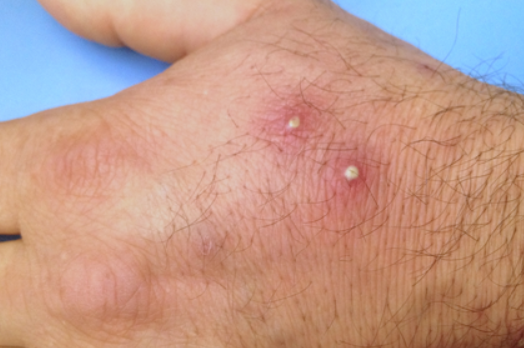
Sterile needles are used to puncture four sites on the forearm. After 24-48 hours, a papule or pustule up to 2 mm in diameter appears at the puncture site and disappears within 3-4 days. This phenomenon is the result of non-specific hyperreactivity. If the test is positive, it has diagnostic value, but its absence does not rule out the diagnosis of Behçet's disease.
The diagnosis of Behçet's disease is based on the classification criteria developed by the International Study Group for Behçet's Disease (ISBD, 1990).
"Major" criteria:
- Recurrent ulcers
- Minor or major aphthae or herpetiform ulcers in the oral cavity, confirmed by a physician or based on reliable patient information, with a recurrence of at least three times per year.
- Recurrent genital ulcers. Aphthae or scars, primarily in males, confirmed by a physician or based on reliable patient information.
- Eye involvement
- Anterior uveitis, posterior uveitis, or cells in the vitreous ( detected by slit-lamp examination).
- Retinal vasculitis (determined by an ophthalmologist).
- Skin involvement
- Erythema nodosum (confirmed by physician or based on reliable patient information)
- Pseudofolliculitis and papulopustular eruptions.
- Acne-like rash (observed in postpubertal patients not receiving glucocorticoids).
- Positive pathergy test.
At least three of the above criteria are required to confirm the diagnosis.
"Minor" criteria.
- Gastrointestinal involvement: Ulcers in the ileocecal region of the intestine.
- Vascular involvement.
- Central nervous system (CNS) involvement.
- Epididymitis.
- Non-deforming and non-ankylosing arthritis.
In several countries, Behçet's disease is classified into different variants:
- Complete type: presence of all four major criteria in a patient.
- Incomplete type: three major criteria, or two major and two minor criteria, or typical ocular symptoms combined with one major or two minor criteria.
- Possible type: only two major criteria or one major and two minor criteria.
This classification approach to Behçet's disease seems reasonable because it contributes to earlier diagnosis of the disease.
In the medical history, needs to pay attention to:
- The presence of recurrent cases of the disease in the families of patients (familial aggregation).
- Episodes of aphthous stomatitis (since childhood).
- Episodes of sudden deterioration of vision or "blurry" vision.
- Visits to a urologist due to scrotal edema.
- Digestive disorders (diarrhea with blood).
- History of thrombophlebitis, primarily in the veins of the lower extremities (more common in males).
- Presence of any cerebral symptoms in the medical history, such as epileptiform seizures.
- Herpes simplex virus
- Pemphigus vulgaris
- Reactive arthritis
- Crohn's disease
- Ulcerative colitis
- Erythema nodosum
- Erosive lichen planus
- Syphilis
- HIV infection
- Aphthous stomatitis
The main goal of treatment is to achieve remission or reduce the number of disease recurrences.
1. Any patient with Behçet's disease with inflammatory involvement of the eye:
- Azathioprine
- Systemic glucocorticoids
2. Severe eye involvement with loss of 2 lines of visual acuity on a 10/10 scale or retinal involvement (retinal vasculitis or macular involvement):
- Cyclosporine A (2-5 mg/kg/day)
- Infliximab + azathioprine and glucocorticoids
3. There is no clear evidence regarding the treatment of large vessel pathology.
- For acute deep vein thrombosis: immunosuppressive agents (glucocorticoids, azathioprine, cyclophosphamide, or cyclosporine A)
- For aneurysms: cyclophosphamide and glucocorticoids
4. There is no evidence that anticoagulants, antiplatelet agents, or fibrinolytics are effective in treating deep vein thrombosis or arterial damage in Behçet's disease.
5. There is no evidence for the treatment of gastrointestinal manifestations of Behçet's disease. The following medications may be prescribed before surgery:
- Sulfasalazine
- Glucocorticoids
- Azathioprine
- TNF inhibitors
6. For arthritis, colchicine (1-2 mg/day) may be used.
7. In cases of central nervous system (CNS) involvement (no controlled studies available):
- Parenchymal involvement:
- Glucocorticoids
- Interferon-alpha
- Dural sinus thrombosis:
- Glucocorticoids
- Azathioprine
- Cyclophosphamide
- TNF inhibitors
8. Cyclosporine A is not used in patients with CNS involvement of Behcet's disease unless intraocular inflammation requires it.
9. Treatment of skin and mucosal manifestations depends on their severity:
- For isolated oral or genital ulcers, local application of glucocorticoids is recommended.
- For erythema nodosum, colchicine may be used.
- For acne-like eruptions, cosmetic measures can be taken.
- In resistant cases, azathioprine and TNF inhibitors may be considered.

