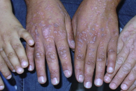
Epidermodysplasia verruciformis (Lewandowsky-Lutz dysplasia) is a rare inherited disorder characterized by widespread wart-like eruptions that tend to become malignant. ICD-10 code is B07.
Epidermodysplasia verruciformis is an autosomal recessive disorder in which approximately 10% of patients are children of affected parents and 30% of siblings are affected. The disease manifests in early childhood. The underlying factors of the disease are:
- Mutations in the EVER1 and EVER2 genes on chromosome 17q25 that prevent the body from preventing and fighting human papillomavirus (HPV) infection.
- Infection with HPV types 3, 5, 8, 9, 10, 12, 14, 15, 17, 19, 20, 21, 22, 23, 24, 25, 28, 29, 36, 46, 47, 49, and 50.
In rare cases, the disease may be acquired rather than inherited in individuals with severe immunodeficiency, such as those with HIV infection, after immunosuppressive therapy following organ transplantation, or in other immunodeficient states. In these cases, the disease can manifest at any age, but is more common in adults.
The disease is characterized by two types of eruptions:
Wart-like eruptions: Multiple (20 or more) widespread, slightly raised papules 3-5 mm in diameter with polygonal borders. They may be flesh-colored, reddish-brown, or dark red, with a smooth or scaly surface. They tend to cluster, have linear arrangements, and coalesce into wart-like plaques. These eruptions are localized on sun-exposed areas of the body, such as the face, shins, and forearms. When localized on the backs of the hands, they resemble flat warts. This type of eruption, especially when caused by HPV types 5, 8, and 14 in individuals aged 30-40 years, is prone to progress to Bowen's disease, Bowenoid papulosis, basal cell carcinoma, or squamous cell carcinoma in 50% of cases.
Hypo- and hyperpigmented spots and patches: Large hypo- and hyperpigmented flat patches with mild scaling resembling multicolored lichen and ichthyosis. They coalesce and are found on the trunk, neck, shoulders, and thighs. Occasionally, tan pigmented areas resembling freckles may appear on the face. These lesions do not tend to become malignant.
Rarely, limited areas of hyperkeratosis, dyschromia, and alopecia may occur. Patients are not considered contagious to immunocompetent individuals.
- Flat warts
- Acrokeratosis verruciformis of Hopf
- Darier's disease
- Actinic keratosis
- Tinea versicolor
- Squamous cell carcinoma
- Basal cell carcinoma
- Molluscum contagiosum
- WHIM syndrome (warts, hypogammaglobulinemia, infections, and myelokathexis)
- WILD syndrome (warts, immunodeficiency, lymphedema, and anogenital dysplasia syndrome)
- Netherton syndrome
Wart-like elements are removed surgically, by cryotherapy, electrocautery, photodynamic therapy, or laser, but recurrences may occur with all of these methods of removal. Systemic and topical retinoid therapy (e.g., oral isotretinoin at a dose of 1 mg/kg/day) can reduce the manifestations of the disease with temporary success. Improvement has been reported with the use of topical imiquimod, tacalcitol, 5% fluorouracil cream and 30% podophyllin ointment, as well as preparations containing salicylic acid and urea. Granulocyte-macrophage colony-stimulating factor, cimetidine, cidofovir, and interferon have been used with variable success.
Preventing eruptions from becoming malignant is important. Prevention includes:
- Regular examination by a dermatologist
- Limiting sun exposure and using sunscreen and protective clothing
- Vaccination against HPV is not recommended for prevention, as it does not protect against the HPV types that cause the disease.

