Congenital dermal melanocytosis (Mongolian spot)

Mongolian spot

Congenital dermal melanocytosis includes pigmented lesions composed of intradermal melanocytes that have not completed migration from the neural crest to the dermo-epidermal junction during fetal life. ICD-10 Code: D22.9

Mongolian spot


Mongolian spot 2
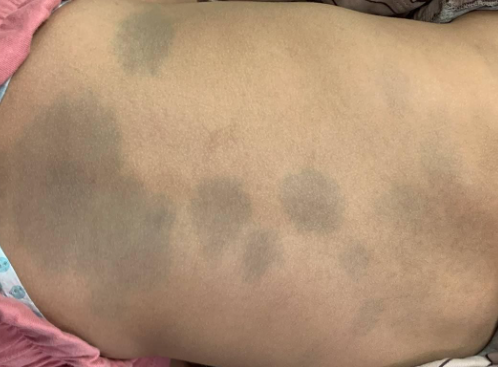
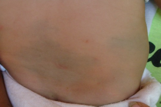
Mongolian spot is a congenital, oval or round, bluish-gray patch up to 10 cm in diameter. It is most commonly located in the sacral-lumbar region. It is observed in 90% of newborns of Asian and American origin and in about 1% of children of European race. The birthmark almost always disappears in early childhood.

Nevus of Ota


nevus of ota
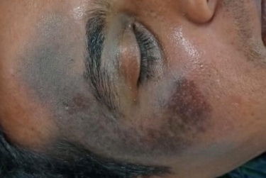
Nevus of Ota is a mottled patch of uneven coloration: diffuse blue pigmentation and brown dots in a reticular or geographic pattern. It is characterized by persistent diffuse pigmentation of the skin around the eyes and sclera caused by accumulation of melanocytes in the dermis. It is usually located in the area of the first and second branches of the trigeminal nerve. This nevus is relatively common in individuals of Asian origin, but rarely affects individuals of other races. Typically, the nevus appears in childhood or during puberty and persists throughout life. There have been reports of malignant melanoma arising from a nevus of Ota.

Nevus Ito


nevus of ito
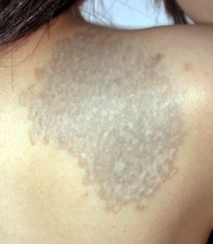
Nevus Ito has a mottled appearance with bluish and brownish spots and is located in the area of the clavicle and upper chest. It is similar to Nevus of Ota but differs in location. The nevus almost exclusively affects individuals of Japanese descent. Malignant transformation is extremely rare.

Diagnosis of an intradermal melanocytic nevus is established clinically and may be confirmed by histological examination.

  • Blue nevus
  • Melasma
  • CafĂ©-au-lait spot
  • Speckled lentiginous nevus (Nevus spilus)
  • Bruise
  • Argyria
  • Ochronosis
  • Drug eruptions
  • Lentigo maligna

Camouflaging with matte makeup reduces the cosmetic defect.

Various procedures such as cryotherapy, dermabrasion, and electrodessication have been used, but these treatments are often associated with hypopigmentation or scarring. Satisfactory results for Nevus of Ota have been achieved through sequential combined application of dry ice, superficial peeling, and argon laser. Excellent cosmetic results have been observed with laser therapy, especially ruby laser with pulse modulation. Alexandrite laser with pulse modulation is also considered a highly effective and safe tool for the treatment of Nevus of Ota.

Long-term follow-up is necessary due to the potential risk of melanoma development, especially in the case of nevi located around the eyes.