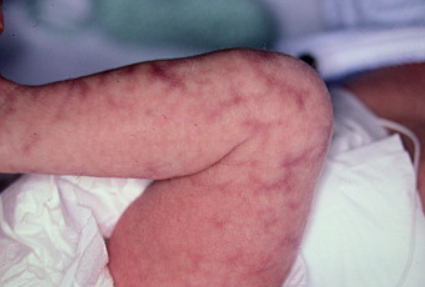
Cutis marmorata telangiectatica congenita is a form of congenital mixed venous-capillary malformation characterized by a reticular (net-like) structure. ICD-10 code: Q27.8
The majority of cases are sporadic, but familial cases have been documented. The male to female ratio is 1:2. It is almost always present at birth (in 90-95% of cases).Localized or regional forms of the disease involving one or two areas of the skin (most often the limbs, less often the face, trunk and neck) are more common than the diffuse type, which affects the entire skin surface. Asymmetric - unilateral or segmental - distribution of lesions is typical. The affected skin areas show fixed livedoid reddish-blue or bluish-violet patches, along with telangiectasias, giving the lesions a "net-like" appearance. The color of the lesions becomes more pronounced with physical exertion, crying, or screaming.
Congenital telangiectatic marbled skin is often associated with "port-wine stains," spider and cherry angiomas, which become more prominent with age as the reticular abnormalities fade.
Atrophy and ulceration may occur already in the neonatal period. The atrophic reticular pattern differs from physiologic marbled skin because of its marked atrophy and irregularity. Involvement of the limbs may lead to atrophy of the subcutaneous tissue and muscles, resulting in reduced volume compared to the healthy limb. Rarely, hypertrophy (pseudoathletic appearance) of the affected limb is observed. Superficial erosions and ulcers often develop in the affected areas, especially around the joints, leaving scars after healing.
In almost half of cases, cutaneous anomalies are associated with musculoskeletal anomalies (syndactyly, micrognathia, high palate, dystrophic teeth, triangular and asymmetrical skull shape), arterial stenosis and heart defects, glaucoma and other eye anomalies, imperforate anus, genital anomalies, congenital hypothyroidism, and others. Cutis marmorata telangiectatica congenita , syndactyly, heart defects and alopecia are integral parts of Adams-Oliver syndrome.
- Macrocephaly-capillary malformation syndrome
- Klippel-Trenaunay syndrome
- Sturge-Weber-Krabbe syndrome
- Mottled skin (livedo reticularis)
- Neonatal lupus.

