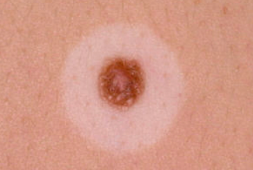
Halo nevus is a melanocytic nevus surrounded by a depigmented zone in the form of a halo. According to the latest concept, it is not considered as a separate nosological entity, but as a phenomenon of halo, inherent in various types of nevi. ICD-10 code: D22.9
It most commonly affects adolescents and young adults, with a general prevalence of 1% in individuals younger than 20 years of age. Familial cases are not uncommon. 18-26% of patients have vitiligo. In addition, halo nevus may be a precursor of vitiligo.
The melanocytic nevus, which often regresses spontaneously, may be a common acquired nevus (intradermal or compound), a congenital or atypical melanocytic nevus, a Spitz nevus, or a blue nevus.
Halo nevus is the result of an immune response (both cellular and humoral) directed either against antigenically altered nevus cells undergoing dysplastic changes or against non-specifically altered nevus cells in response to various insults. Immune mechanisms are involved in the process of central nevus regression. Cytokines may act as mediators in the development of the white halo.
Halo nevus clinically presents as a dark brown or pink color central macule or papule with a well-defined border of regular shape surrounded by a symmetric, circular or oval, hypopigmented or white ring that is clearly demarcated from the surrounding skin. The lesion causes no symptoms and may be associated with mild pruritus. It most commonly occurs on the trunk, especially the back, but can affect any area of the skin. Multiple lesions occur in 25-50% of patients.
Halo develops over months around a pre-existing melanocytic nevus. The course of the disease is variable. Rarely does the lesion remain unchanged. More commonly, the central nevus regresses spontaneously, followed by repigmentation of the white halo. This process may take months to years. In some cases, the depigmented spot remains, while in others the halo re-pigments and the central nevus remains unchanged.
Malignant transformation can complicate halo nevus. Occasionally, halo may form around a primary malignant melanoma. In older adults, the unexpected appearance of multiple halo nevi is associated with the development of a primary cutaneous malignant melanoma elsewhere.
- Nevus Spitz
- Blue nevus
- Congenital melanocytic nevus
- Dysplastic nevus
An asymmetrical and irregularly shaped halo surrounding a large lesion with pronounced color variation may indicate the presence of the halo phenomenon in primary cutaneous malignant melanoma.
Halo nevi with a benign clinical appearance do not require excision. The patient should be reassured and periodically monitored. Lesions with atypical clinical features should be excised for histological examination. Individuals over 40 years old, as well as patients with a family history of skin melanoma or dysplastic nevi, should be thoroughly evaluated for the presence of cutaneous malignant melanoma.

