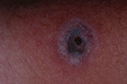
Gangrenous ecthyma (ecthyma gangrenosum) is a skin infection, most commonly caused by Pseudomonas aeruginosa bacteria, characterized by the formation of necrotic lesions covered by a crust. ICD-10 code L08.0.
Gangrenous ecthyma is a rare disease that can affect people of all ages, ethnicities, and genders. It is caused by strains of Pseudomonas aeruginosa. The exact pathogenesis remains unclear. According to some investigators, there is an initial skin infection followed by sepsis due to hematogenous spread of the infection, while others consider ecthyma to be a secondary process in cases of already developed pseudomonal sepsis. Infection can enter the body through skin wounds, insect bites, burns, pressure ulcers, injections, tracheal surgery, urological procedures, catheterization of blood vessels and the urinary bladder.
Gangrenous ecthyma typically develops in patients with weakened immune systems, including those with hematologic malignancies, acquired and congenital immunodeficiency syndromes, neutropenia, pancytopenia, severe burns, malnutrition, individuals undergoing chemotherapy and immunosuppressive therapy, and those with diabetes mellitus. The use of antibiotic treatment to which Pseudomonas is resistant may result in its active proliferation.
However, cases of the disease have been reported in individuals with normal immunity and in previously healthy individuals. In some cases, gangrenous ecthyma may be caused by other microorganisms. In such cases, it is referred to as non-pseudomonal gangrenous ecthyma or ecthyma-like lesions. The following etiologic classification has been proposed:
- Pseudomonas-associated gangrenous ecthyma (Pseudomonas aeruginosa, Pseudomonas cepacia, Pseudomonas maltophilia, and Pseudomonas stutzeri)
- Non-pseudomonal gangrenous ecthyma includes:
- Bacterial infections in immunocompromised and immunocompetent patients. (Staphylococcus aureus,Streptococcus pyogenes,Aeromonas hydrophila,Klebsiella pneumonia,Serratia marcescens,Xanthomonas maltophilia,Morganella morganii,Escherichia coli,Citrobacter freundii,Corynebacterium diptheriae,Neisseria gonorrhea,Yersinia pestis,Stenotrophomonas maltophilia)
- Fungal infections with or without septicemia. (Candida species,Aspergillus fumigatus,Aspergillus niger,Fusarium solani,Scytalidium dimidiatum,Pseudallescheria boydii,Curvularia species,Meiarrhizium anisopliae,Mucor species,Exserohilium species)
- Viral infections. (Herpes simplex virus)
The disease typically begins with the formation of a painless erythematous spot or petechiae, which becomes painful within 12-24 hours and transforms into a hemorrhagic vesicle or blister. Within a day, the lesion becomes infiltrated and the vesicle or bulla ruptures, forming an ulcer with a black necrotic crust on its surface. An erythematous ring is observed around the periphery of the lesion. The affected area grows rapidly, sometimes reaching 10 centimeters or more in diameter. The rash is most commonly found on the axillary folds, groin, and perineum (apocrine zones), and less commonly on the buttocks, trunk, and limbs. The face is rarely affected. If left untreated, it can lead to pseudomonal sepsis with fever, hypotension, chills, and diarrhea. There may be a fine pink rash on the trunk, petechiae, ecchymosis, and clusters of painful vesicles and bullae with subsequent formation of black crusts. The mortality rate varies from 18% to 96%, mainly due to disseminated intravascular coagulation.
Clinical Variants:
- Noma neonatorum occurs in premature infants. Besides the formation of lesions in the anogenital area, the face and oral cavity are frequently affected.
- Necrotizing stomatitis - a manifestation of gangrenous ecthyma on the mucous membrane of the oral cavity, also observed in severely immunosuppressed adult patients.
- Herpes simplex
- Antiphospholipid syndrome
- Polyarteritis nodosa
- Cryoglobulinemia
- Cryofibrinogenemia
- Cellulitis
- Ecthyma
- Siberian ulcer
- Necrotizing fasciitis
- Gas gangrene
- Acute meningococcemia
- Calciphylaxis
Treatment should be aimed at complete eradication of the pathogen. However, broad-spectrum antibiotics are usually used at the beginning of therapy because there is often no time to accurately identify the microorganism and its susceptibility to antimicrobial agents. Delayed initiation of antibiotic therapy may result in increased patient mortality. The prognosis of the disease depends on the type of pathogen, the duration and severity of immunosuppression, and the extent of skin involvement.
If gangrenous ecthyma is suspected before the results of microbiologic testing are available, urgent antibiotic therapy is initiated with a combination of aminoglycosides (tobramycin, gentamicin, or amikacin) and penicillins (carbenicillin, piperacillin, or ticarcillin), cephalosporins (ceftriaxone or cefepime), or carbapenems (meropenem).
Recommended antibiotic dosages:
- Ceftazidime: 2 g intravenously every 8 hours
- Cefepime: 2 g intravenously every 12 hours
- Ticarcillin: 3 g intravenously every 4 hours
- Gentamicin or Tobramycin: 3-5 mg/kg intravenously 2-3 times daily
- Meropenem: 0.5 g every 8 hours or imipenem: 0.5 g every 6 hours
- Amikacin: 7.5 mg/kg intravenously every 12 hours

