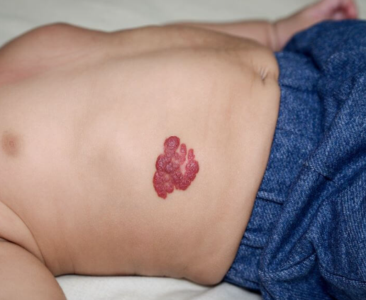
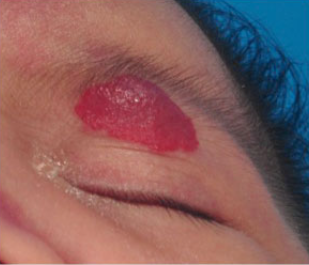
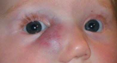
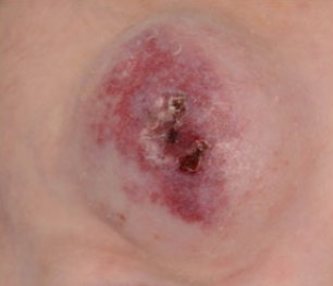
Infantile hemangiomas are benign vascular tumors with signs of cell proliferation and a typical pattern of development: a period of active growth is followed by stabilization and subsequent involution. ICD-10 code: D18.0.
The vast majority of them are benign tumors of infancy, occurring in 1.0-2.6% of newborns. Their frequency in children under 1 year of age is about 10-12% in the white population. They are particularly common in premature infants, and there is a close correlation between the frequency of hemangiomas and the birth weight of the child (approximately 1 in 4 newborns with a weight of less than 1000 grams has a hemangioma), although a possible correlation with gestational age cannot be excluded. Hemangiomas are 4-6 times more common in girls than in boys.
The exact etiology and pathogenesis of hemangiomas are unknown because they involve numerous cellular and molecular components. Some molecules, including proliferating cell nuclear antigen, collagenase IV, vascular endothelial growth factor, and basic fibroblast growth factor, are highly expressed on endothelial cells. These molecules partially mediate endothelial cell differentiation and then attract adipocytes during the rapid growth phase of the hemangioma. Late expression of a potent inhibitor of new blood vessel formation - tissue inhibitor of metalloproteinase type 1 - may be an important factor contributing to the involution phase of hemangiomas.
Depending on the clinical and histologic presentation, infantile hemangiomas can be classified into the following types:
- Superficial (also known as strawberry hemangiomas), which account for 50-60% of cases.
- Deep (previously called cavernous hemangiomas), which account for 15% of cases.
- Mixed types, which make up 25-30% of all cases.
Superficial hemangiomas
Deep hemangiomas
Mixed hemangiomas
Course of hemangiomas:
Regression of deeper lesions is more challenging to detect, but the time and rate of the process are equivalent. Approximately 40% of patients retain residual skin changes after involution, including scarring, atrophy, excess skin, altered pigmentation, and telangiectasias. Approximately 50% of lesions are located on the head and neck, 35% on the trunk, and the perianal area is also a favored site. Hemangiomas in children may be solitary or multiple. Approximately half of the hemangiomas are present at birth, while the remainder typically appear in the first few months of life. The characteristic growth phase follows the initial appearance of the lesions at approximately 6-10 months, although deep hemangiomas may continue to grow slowly for several months. The lesions usually reach their maximum size by the end of the first year of life. Most regress spontaneously within an average of 2-6 years.
Complications:
- Trauma or erosion of lesions, especially larger ones, may cause bleeding.
- During the rapid proliferative phase, there is a tendency for ulceration, especially in the anogenital area and traumatically exposed areas such as the ears, nose or lips.
- A rare complication is heart failure, which is usually associated with large or multiple hemangiomas.
- Systemic hemangiomas are a sign of multiple organ involvement, and extensive systemic lesions can lead to high mortality.
- Kasabach-Merritt syndrome is characterized by consumption coagulopathy associated with a large deep hemangioma or, rarely, multiple small hemangiomas or angiomas in internal organs.
- Eyelid hemangiomas may cause visual impairment due to possible obstructive amblyopia or astigmatism.
- Sublingual hemangiomas may cause airway obstruction, especially if the cutaneous lesion is located on the neck.
- Obstruction of the external auditory canal by hemangiomas can rapidly lead to hearing loss.
- Direct pressure from the tumor can lead to deformation of the underlying bone tissue.
- Vascular malformations
- Pyogenic granuloma
- Myofibromatosis
- Spitz nevus
- Dermoid cysts
Since hemangiomas are benign in nature and usually resolve on their own, it is best not to intervene in most cases.
Parents should be reassured about the good prognosis and advised to visit the doctor regularly. Photographing the tumor at regular intervals can document changes in the lesions during follow-up. Measures should be taken to provide local care for the lesions to prevent ulceration and secondary infection, especially for lesions in the anogenital area.
Commonly used treatments include antibiotics, barrier creams, and biocellulose dressings.
A small number of patients may require treatment for the following lesions:
- Life-threatening and functionally significant hemangiomas (e.g., hemangiomas that cause visual impairment, interfere with feeding, lead to airway obstruction, or cause Kasabach-Merritt syndrome and heart failure).
- Hemangiomas in specific anatomic areas that may cause permanent scarring or deformity (e.g., lesions on the nose, lips, ears, or forehead).
- Large facial hemangiomas, especially those with a prominent dermal component, which tend to leave permanent scarring.
Treatment options include:
- Corticosteroids (systemic, intralesional, and topical).
- Interferon-alpha therapy.
- Laser therapy.
- Cryosurgery.
- Surgical excision.
- Compression under occlusion.
- Sclerotherapy.
- Embolization.


