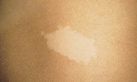
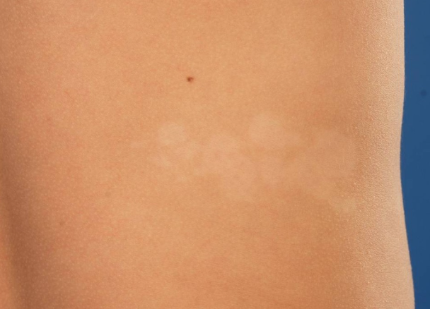
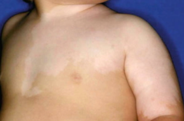
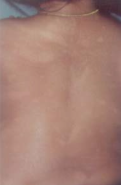
Nevus depigmentosus, a congenital skin disorder, is characterized by a localized hypopigmented spot that is present from birth or appears in early infancy. ICD-10 code is L81.8.
The disease affects both sexes and all ethnic groups. In most cases, it is detected at birth, but it can also appear within the first three years of life. The prevalence of the condition is unknown.
The exact causes and mechanisms of nevus depigmentosus are not fully understood. It is thought to be related to functional abnormalities in the transfer of melanosomes from melanocytes to keratinocytes, resulting in the development of characteristic hypopigmented spots of skin. Some researchers suggest that nevus depigmentosus may be a form of cutaneous mosaicism in which altered clones of melanocytes have a reduced capacity to synthesize and transport melanin to keratinocytes. The presence of lentigines alongside nevus depigmentosus may also represent a phenotypic manifestation of reverse mutation.
Some researchers consider nevus depigmentosus and hypomelanosis of Ito to be two phenotypic variants of mosaicism affecting pigment genes. The genetic heterogeneity may be related to multiple loci encoding pigment-related proteins. In nevus depigmentosus, somatic mutations occur late during embryogenesis when most structures are already formed, resulting in fewer systemic manifestations. In contrast, in hypomelanosis of Ito, these mutations are likely to occur much earlier, increasing the likelihood of systemic involvement (62-94%).
Localized form
Segmental form
Systemic form
Disease progression
The hypopigmented spots are more noticeable on darker skin and become more pronounced with tanning. The disease is chronic and persistent. The size of the spots tends to increase with age, but usually stabilizes after puberty. Isolated cases of spontaneous regression of the disease have been reported. In the absence of systemic manifestations, nevus depigmentosus is considered benign.
Clinical diagnostic criteria (Coup, 1976):
- The hypopigmented spot is present from birth or appears in early infancy.
- There are no changes in the distribution of the lesion throughout life.
- There are no changes in texture or sensation in the affected area.
- Absence of a hyperpigmented border.
Clinical investigations:
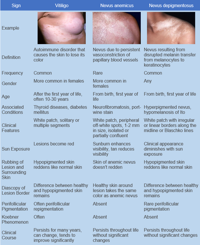
Diascopy and rubbing test are negative, ruling out anemic nevus.
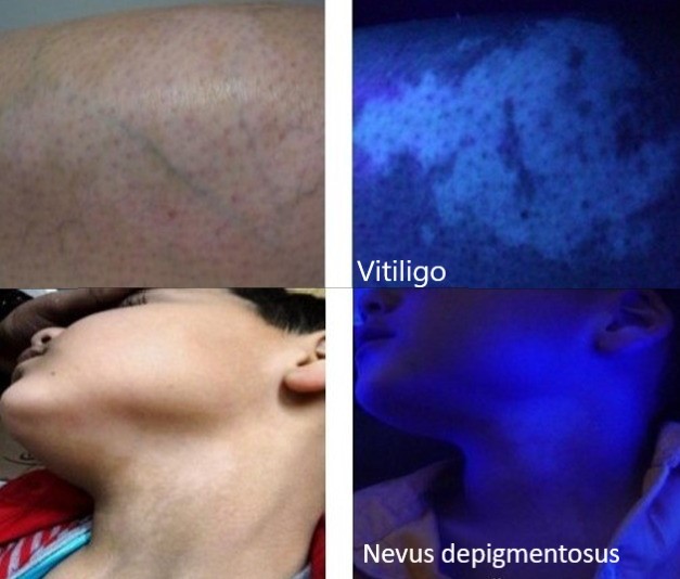
Wood's lamp examination: In contrast to vitiligo, there is a whitish fluorescence with clearer demarcation of the lesion, but the border with healthy skin appears blurred.
Treatment is not required. The following is recommended for cosmetic purposes:
- Camouflage (especially for exposed body parts)
- PUVA therapy
- Excimer laser
- Transplantation of melanocytes

