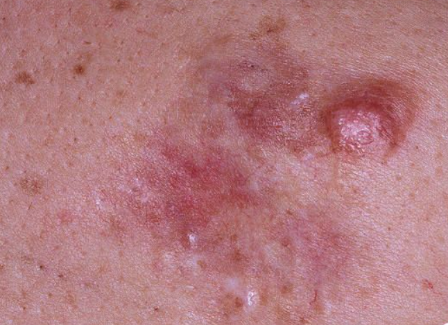
Dermatofibrosarcoma protuberans is a connective tissue tumor with infiltrative growth and a tendency to recur. ICD-10 Code: C49.9.
It is more often observed in men between 30 and 40 years of age, and in about 10% of cases it occurs in children. Most authors consider fibroblasts with active collagen synthesis in a well-developed endoplasmic reticulum as the main cells of the tumor. The cells have spindle-shaped nuclei, and some of them are surrounded by material resembling interrupted basal membranes, suggesting their possible origin from perineural or endoneural elements.
In addition to the main variant, an atrophic (morphea-like) variant of the tumor and a pigmented form (Bednar's tumor) have been described.
Dermatofibrosarcoma protuberans typically occurs on the skin of the trunk, extremities, neck, and rarely on the scalp or genitalia. It usually presents as a solitary tumor, but may consist of multiple morphea or keloid-like nodules over a relatively large area (up to 20 cm in diameter). Such a plaque may have a rounded or polycyclic shape, distinct borders, and a yellowish-white, purplish, or reddish-brown color, visibly protruding from the surrounding skin. Gradually, several round and firm tumors develop on this background and reach a large size (sometimes as big as a child's head). At first, the new growth is not attached to the underlying tissue, but later it tends to infiltrate the subcutaneous tissue, muscles and fascia. Sometimes it may erode due to friction with clothing, but ulceration is rare. The tumor usually progresses without subjective sensation, although pain may be present in some cases. An atrophic (scleroderma-like) variant of the tumor has also been described.
The disease progresses slowly, with rare exceptions when rapid growth occurs, sometimes with the formation of satellite nodes or nodules (during pregnancy). Metastasis is observed in about 4-5% of patients, but after many years of tumor existence. Hematogenous metastases (75%) are more commonly found in the lungs, while lymphogenous metastases (25%) are observed in regional lymph nodes.
Dermatofibrosarcoma protuberans is characterized by a high recurrence rate after removal (even many years later). The prognosis is serious, although better than for true sarcomas. Cases of tumor development in HIV-infected individuals and complex tumors including giant cell fibroblastoma and endometriosis have been described.
Bednar's tumor (Pigmented Dermatofibrosarcoma Protuberans, Fibrosarcomatous Bednar's Tumor) is a very rare variant (about 5%) of Dari-Ferrand's tumor and is clinically almost indistinguishable from the latter. It has a brown or dark brown color and is often multilobular. Metastases, in some cases multiple, rarely occur, mainly in the lung, sigmoid colon, chest wall, supraclavicular region and parafaringeal.
- Fibrosarcoma
- Dermatofibroma
- Diffuse type neurofibroma
- Leiomyosarcoma
- Melanoma
- Pilomatrixoma
- Giant cell fibrosarcoma
Radical surgical excision is necessary.
Prognosis is relatively favorable.

