Leukoplakia

leukoplakia

Leukoplakia is a dystrophic change of the oral mucosa accompanied by varying degrees of epithelial hyperkeratosis and is classified as a precancerous condition. ICD-10 Code: K13.2

Leukoplakia most often develops between the ages of 40 and 70. Men are affected twice as often as women. It is one of the most common precancerous lesions of the oral cavity, observed in 0.2-4.9% of the population.

Tobacco use plays an important role in its pathogenesis (80-90% of patients are smokers). There is a synergistic effect with alcohol consumption. Chronic traumatization of the mucous membrane by dentures and sharp edges of teeth is also an important factor.

The role of Sanguinaria extracts, present in some toothpastes and mouthwashes, has been studied. Since leukoplakia often coexists with Candida fungal infection, the possible effect of such infection on malignant transformation has been studied. Certain fungal strains have been found to produce nitrosamine, a known carcinogen.

The involvement of Epstein-Barr virus in disease development is not proven, but human papillomaviruses (HPV-16 and 18) may in some cases contribute to the development of leukoplakia and its malignant transformation.

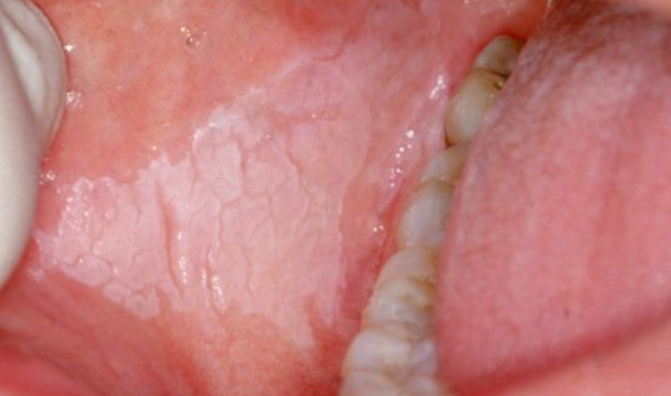
Flat type


flat leukoplakia
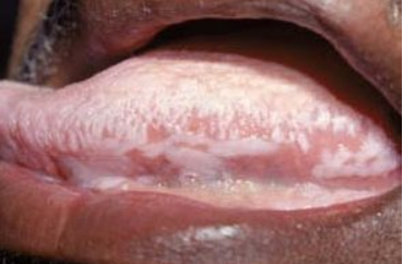
Characterized by the appearance of milky white patches of various sizes and shapes with well-defined borders on the mucous membranes. These patches are not elevated above the mucosal surface and are covered with a multilayered flat epithelium with areas of difficult to remove keratosis. There are no subjective symptoms.

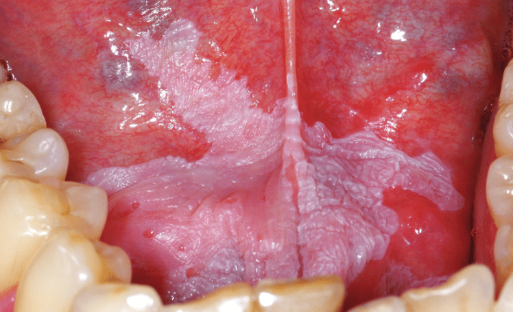
Verrucous type


Verrucous leukoplakia
Presented by plaque-like lesions with wart-like surfaces and prominent keratosis. These lesions are pale white, slightly dense, and protrude above the surrounding mucosa. Sometimes they are found next to areas of flat leukoplakia. There are no subjective symptoms.

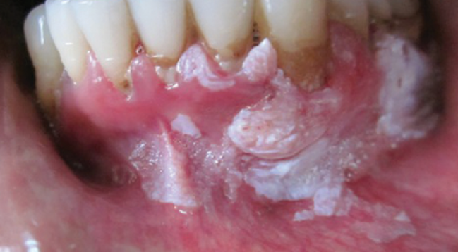
Erythroleukoplakia


Erythroleukoplakia
Represents a combination of mucosal lesions characterized by both erythroplasia of Queyrat and homogeneous leukoplakia, appearing as patches and spots of red and white color. There are variants with predominance of either the first or second component. Patients experience pain, pruritus, discomfort, and dysphagia. Predominant localization includes the soft palate, floor of the mouth, cheeks, and tongue.

Hairy Leukoplakia


Hairy Leukoplakia
Almost exclusively observed in HIV-infected individuals. This condition has been shown to result from infection of the mucosa by the Epstein-Barr virus and/or HPV. In some cases, "hairy" leukoplakia is found in other immunodeficient conditions and exceptionally rarely in healthy individuals. It manifests as whitish patches with a rough "hairy" surface or a smoother surface with vertical fissures.

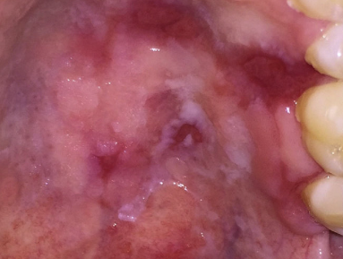
Nicotine stomatitis


Nicotine stomatitis
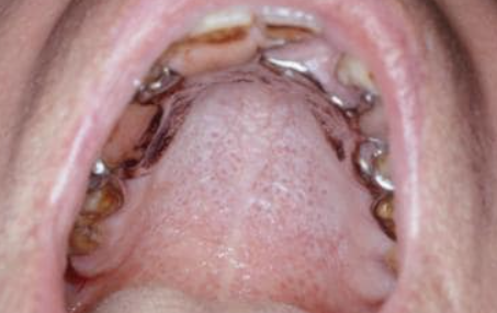
Nicotine stomatitis is mainly localized on the hard palate of smokers, especially those who use pipes. Small hemispherical nodules of the same color are seen against the grayish white mucosa. The nodules have gaping openings of salivary gland ducts on their tops, which appear as reddish dots. The closely spaced papules, with folds forming between them, give the focus the appearance of a cobblestone pavement.

Diagnosis is based on clinical data and biopsy results.

  • Psoriasis
  • Lupus erythematosus
  • Lichen planus
  • Candidiasis
  • White sponge nevus
  • Secondary syphilis

  • Elimination of traumatizing factors in the oral cavity and smoking.
  • Surgical excision, cryotherapy, laser ablation are indicated.
  • Topical corticosteroids are also prescribed, such as clobetasol ointment, applied topically 3 times/day for the first month, 2 times/day for the second month, and 1 time/day for the third month.
  • Methods of combined topical treatment with retinoids in combination with interferon administration have also been described.
  • Phototherapy.

Prognosis: Squamous cell carcinoma develops in 2-4% of cases.