Leiomyoma is a benign tumor composed of smooth muscle cells. ICD-10 Code: D21.9
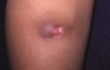
Angioleiomyoma is a benign tumor that develops from the muscular elements of small blood vessels in the skin. ICD-10 Code: D21.9
Leiomyosarcoma is a malignant tumor that arises primarily from the muscles associated with hair follicles. ICD-10 Code: C49.9
Myogenic tumors account for approximately 10% of all soft tissue neoplasms of the skin. The ratio of smooth muscle tumors to striated muscle neoplasms is 100:1. Piloleiomyomas account for approximately 10% of all cutaneous leiomyomas. Multiple forms are more common than solitary. The female to male ratio is 2:1, increasing to 8:1 in familial cases and 5:1 in sporadic cases.
The exact etiopathogenesis is unknown.
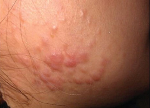
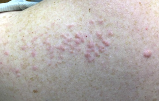
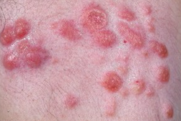
Multiple Leiomyomas
Leiomyomas are characterized by dense, elevated, oval papules ranging in size from a pinhead to a bean. Their color varies from reddish to bluish-red to normal skin color, and they have a smooth, shiny surface. The nodules are arranged in groups, segments, or linear patterns: more often on the head, trunk, and lower limbs in men, and more often on the legs in women. In 73% of cases, the rash is unilateral. The tumors grow slowly, reaching a diameter of 0.3-1.5 cm.
They are typically painful to touch, with mechanical irritation and exposure to cold causing discomfort. Some authors believe that painful sensations appear in the later stages of development.
Pseudo-Darier sign - Rubbing, exposure to cold, or electric current causes temporary elevation, enlargement, redness, and/or piloerection of the tumor.
Virtually all patients experience pain crises, usually in the form of paroxysms, lasting from a few minutes to 1.5-2 hours. They are often accompanied by a drop in blood pressure, dilation of the pupils, pallor of the skin, vomiting, and involuntary urination.
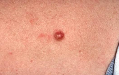
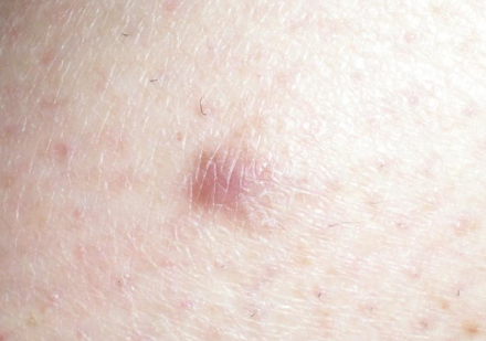
Solitary leiomyoma
Solitary leiomyoma presents as a dense, painful, reddish flesh-colored lump 2 cm or larger in diameter. It can appear on various parts of the body, but is more common on the upper extremities. Small erosions are often found on the surface.
Pseudo-Darier sign is positive.
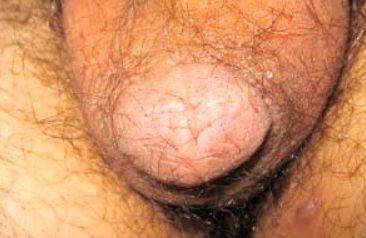
Dartoic leiomyoma
Rare, typically appearing as solitary, painless, dense nodules 1.5-3.0 cm in diameter with a brownish-red color. They are found in the scrotum, labia majora, and rarely on the nipples.
Palpation, exposure to electric current or cold induces worm-like contractions. A zone of mild hyperemia forms around the tumor.
Hereditary Cutaneous Leiomyomatosis (Reed Syndrome)
Hereditary cutaneous leiomyomatosis, also known as Reed syndrome, is characterized by multiple benign skin tumors originating from the muscles associated with hair follicles, uterine leiomyomas, and a high risk of early development of uterine fibroids, female infertility, and renal cancer.
The average age of onset is 20-30 years. Cutaneous leiomyomas are usually small, firm, slightly elevated, oval-shaped growths with a color ranging from normal skin color to yellowish, reddish-brown, or bluish. These multiple elements are often grouped and arranged segmentally or linearly on the body. The lesions appear predominantly on the extensor surfaces of the limbs and buttocks.
The tumors are typically painful to palpation, external stimuli such as temperature changes, psychological stress, or without apparent reason.
In women, there is a hormonal activation of the process during puberty, pregnancy and hormonal contraception. The appearance of skin leiomyomas often precedes the development of uterine fibroids by a significant period of time. Uterine involvement leads to infertility and often requires early hysterectomy at a young age. The disease is slow but progressive. Patients have an increased risk of kidney cancer.
Atypical Leiomyoma
Angioleiomyoma
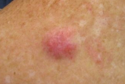
Leiomyosarcoma
Leiomyosarcoma is a rare disease (accounting for 2 to 9% of all soft tissue sarcomas) that occurs equally in both sexes at any age, predominantly in middle-aged and elderly individuals. The tumor is solitary, rarely multiple, with a firm elastic consistency, well defined contours, smooth surface, measuring 1.5-5.0 cm or more in diameter. Nodule is located deep in the dermis and may protrude above the surrounding skin, which takes on a pink or pinkish-blue color. Ulceration of the central zone of the nodule may occur.
The most common sites are the extensor surfaces of the shoulders, forearms, lower limbs, and less commonly the trunk, head, neck, and genitals. The tumor typically doesn't cause subjective sensations, and about 30% of patients experience pain.
The disease is highly aggressive, with frequent metastases to the lungs, liver, and bones. Fatal outcomes can occur within a few months to 3-6 years. Recurrence after surgical removal is observed in the majority of patients. Subcutaneous tumors arising from vascular smooth muscle cells have a worse prognosis than intracutaneous tumors arising from the arrector pili muscles. The former are more likely to metastasize, while the latter tend to recur locally.
- Neurofibromatosis
- Kaposi Sarcoma.
- Neurinoma
- Mastocytoma
- Syringoma
- Eccrine Spiradenoma.
- Glomus Tumor
- Hemangioma
- Surgical excision, electroexcision, laser coagulation (CO2), or cryotherapy for solitary lesions.
- Intralesional injection of botulinum toxin.
- To alleviate pain, α-adrenergic blockers in combination with calcium channel blockers, antidepressants, and analgesics are indicated.
- Nifedipine 10 mg 2-3 times a day.
- Gabapentin 300 mg 2-3 times a day.
- Topical lidocaine spray.
- Leiomyosarcoma - surgical excision with margins.
Prognosis:
Favorable for solitary tumors.
Relatively favorable for multiple tumors.
Unfavorable for leiomyosarcoma.

