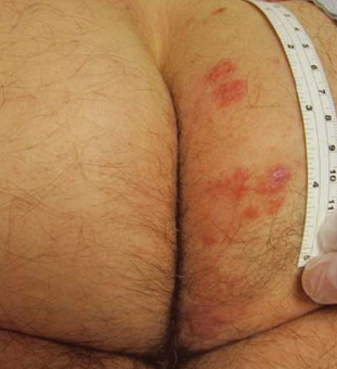
Lymphomatoid contact dermatitis (also known as toxic sofa dermatitis) is a distinct form of allergic, persistent, and chronic contact dermatitis with clinical and/or histopathological features resembling cutaneous lymphoma. ICD-10 code: L98.8.
Lymphomatoid contact dermatitis is more common in men with an average age of 58.5 years. The prevalence is unknown due to underestimation of the condition. This type of contact dermatitis is part of the spectrum of cutaneous lymphomatoid hyperplasia known as pseudolymphoma. The aetiopathogenesis is not fully understood but is thought to involve an immune mechanism of delayed-type hypersensitivity (type IV hypersensitivity reaction) triggered by sensitised T lymphocytes on contact with a specific antigen, leading to reactive lymphoproliferative processes.
The first antigen identified in 1976 was sesquisulfide of phosphorus, which is found in the heads of matchsticks that ignite when rubbed against surfaces. Later cases of the disease were described in association with contact with:
- Organic dyes (diaminodiphenylmethane, paraphenylenediamine, ethylenediamine dihydrochloride, isopropylidenediphenylamine)
- Rubber products (mercaptobenzothiazole)
- Plastics and plywood (paratertiary butyl phenolic resin)
- Metals (gold thiosulfate, nickel sulfate, cobalt naphthenate)
- Preservatives (methylchloroisothiazolinone, quaternium-15)
- Disinfectants (glutaraldehyde)
- Medications (benzydamine hydrochloride)
- Nail lacquers and gels (acrylates)
In 2007, an outbreak of dermatitis on the thighs, legs, and buttocks associated with the mold inhibitor dimethyl fumarate was reported in the UK and Finland. This substance was inserted into leather furniture packages, leading to the term "toxic sofa dermatitis."
Two clinical forms of the disease are recognized: a localized form, which is more common, and a rare generalized form.
Both forms are characterized by pruritus, sometimes severe. The course of the disease is chronic and persistent with continuous exposure to the allergen. It is believed that lymphomatoid contact dermatitis may evolve into a true lymphoma over time.
Localized form:
It is characterized by solitary or multiple flat infiltrated erythematous plaques of round or irregular shape, up to a few centimeters in diameter, and less commonly macules, papules, or nodules of red or purple color. Pigmented lesions have been described. The surface of the plaques may be smooth or covered with gray scales. Occasionally, the rash elements coalesce into larger patches. The rash is usually located on areas of contact with allergens. The most commonly affected areas include the thighs, buttocks, lower trunk, face, hands, and feet. Cases of involvement of the earlobes and genitals have also been described.
Generalized form:
It is characterized by exfoliative erythroderma resembling psoriasis.
Diagnosis is based on the characteristic clinical presentation and histopathological features.
Diagnostic Criteria for lymphomatoid contact dermatitis
- Localized or generalized rash with a history and visual examination suggesting contact dermatitis.
- Histopathological features resembling cutaneous lymphoma.
- Positive patch testing for allergens.
- Rapid regression of eruptions after local corticosteroid therapy and discontinuation of contact with the allergen.
- T-cell and B-cell lymphomas
- Large plaque parapsoriasis
- Lymphomatoid papulosis
- Chronic actinic dermatitis
- Jessner lymphocytic infiltration
- Other Pseudolymphoma
Discontinuation of contact with the allergen and topical corticosteroids result in rapid resolution. Regular follow-up every six months for several years is recommended.

