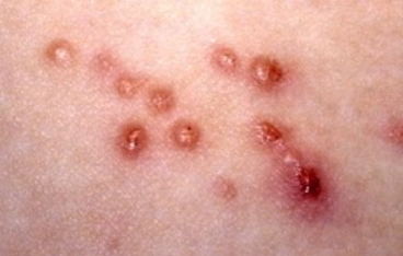
Lymphomatoid papulosis is a chronic recurrent lymphoproliferative skin disorder characterized by recurrent eruptions of spontaneously resolving papular lesions with histologic features resembling lymphoma. ICD-10 Code: D23.9
Lymphomatoid papulosis can occur in all age groups, with a mean age of onset of 35-45 years. The male to female ratio is 1.5:1. The incidence is 1.2-1.9 cases per 1,000,000 population between the ages of 8 and 60.
The etiology and pathogenesis are still unclear. It is thought that papulosis represents an immune response to unknown antigens of relatively low immunogenicity. Some literature presents ultrastructural and clinical evidence supporting a viral etiology. However, conclusive data are lacking, including the role of oncogenic viruses such as HHV-8 and T-lymphotropic HHV-6 and HHV-7. In Asian countries, the disease is often associated with Epstein-Barr virus infection.
Characterized by recurrent eruptions of spontaneously resolving papular lesions. The number of eruptions may vary from a single one to several dozens (sometimes hundreds), and there is a characteristic evolutionary polymorphism of the lesions. The most common localization is on the skin of the trunk and proximal parts of the extremities. Cases of eruptions on the palms and soles, face, scalp, and anogenital area have been described.
The clinical presentation typically consists of rapidly growing asymptomatic red or bluish-red papules up to 2 cm in diameter that persist for three weeks to several months and then resolve completely or ulcerate, leaving hyperpigmented spots or atrophic scars.
In most cases, lymphomatoid papulosis follows a chronic benign course without affecting survival. However, patients are at high risk of developing secondary cutaneous lymphoproliferative disorders, including lymphomatoid papulosis-associated lymphomas such as mycosis fungoides and Hodgkin's lymphoma. These associated lymphomas develop in 10-25% of cases and may precede, occur concurrently with, or follow the onset of lymphomatoid papulosis and must be considered in the diagnostic process.
- Pityriasis lichenoides et varioliformis acuta (PLEVA)
- Papulonecrotic vasculitis
- Nodular prurigo
- Hodgkin lymphoma
- Regressing atypical histiocytosis
- Histiocytosis X
- Lymphomatoid granulomatosis
Due to the heterogeneity and low prevalence of the disease, the number of controlled clinical trials is limited. As a result, all recommendations in this section have a level of evidence C and D. Studies of the effectiveness of various treatments have shown that there is currently no therapy capable of altering the course of the disease or preventing the development of secondary lymphomas associated with lymphomatoid papulosis. Therefore, a strategy of watchful waiting without active therapeutic intervention is preferred.
Treatment Regimens:
For patients with numerous and disseminated eruptions, the best results are achieved with PUVA therapy and low-dose methotrexate treatment (5-30 mg per week with 1-4-week intervals). Both treatments lead to a reduction in the number of eruptions and rapid resolution in the majority of patients. However, complete remission is rare, and relapses quickly occur upon treatment cessation (or dose reduction). Due to the tendency to relapse, maintenance therapy may be required to control the disease progression. It should be noted that prolonged use of PUVA therapy may increase the risk of skin cancer, and methotrexate use may lead to liver fibrosis.
Patients with nodular eruptions > 2 cm in diameter that do not resolve within several months may undergo surgical removal of the lesions or local radiation therapy as an alternative approach instead of the "watch and wait" strategy.
Persistent nodular eruptions > 2 cm in diameter without spontaneous resolution require repeated skin biopsy to exclude secondary anaplastic large cell lymphoma.
Relapses and Subsequent Follow-up:
Patients should be under lifelong surveillance due to the risk of developing secondary lymphoproliferative disorders (4-25% of cases), even decades after disease onset in the absence of skin eruptions. Annual check-ups with chest X-rays and abdominal and pelvic ultrasound are recommended.

