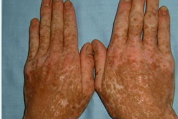
Dyschromatosis symmetrica hereditaria of Dohi is a rare genodermatosis characterized by symmetric widespread reticulated hyperpigmentation of the skin, primarily on the acral areas. The ICD-10 code for this disorder is L81.8.
The condition is found in all countries of the world, but it is most commonly observed in the populations of China and Japan. According to some researchers, the condition is predominantly found in individuals of European descent who have red hair.
The inheritance pattern is autosomal dominant with high penetrance. There are also reports of autosomal recessive inheritance. Rarely, the condition may occur sporadically. The pathogenesis is poorly understood. The disease is primarily caused by a mutation in the ADAR1 gene located at locus 19213.It is characterized by the appearance, usually during the first year of life or later, of eruptions in the form of hypopigmented spots measuring 5-7 mm in diameter on the dorsal surfaces of the hands and feet. Subsequently, areas of hyperpigmentation with a speckled, reticular, or less commonly linear pattern develop. The rash may later spread to the proximal areas of the extensor surfaces of the limbs, more commonly affecting the lower limbs. The palms and soles are usually not affected. Approximately 50% of patients may have isolated eruptions on the face resembling freckles.
On the skin of the limbs, brownish pigmented spots of irregular shape alternate with areas of hypopigmentation, giving the affected areas a reticular appearance. The number of eruptions gradually increases until adolescence and remains stable throughout life.
Skin eruptions can be associated with intellectual disability, neurological disorders, retinal vessel dilation, dystonia, and acral hypertrophy. Patients with this condition have an increased frequency of psoriasis occurrence and a risk of developing epithelial and melanocytic skin neoplasms.- Reticulate acropigmentation of Kitamura
- Dyschromatosis universalis hereditaria (DUH)
- Xeroderma pigmentosum
- Dowling-Degos disease (reticulated pigmented anomaly of flexures)
- Amyloidosis cutis dyschromica
- Vitiligo
- Contact or drug-induced leukoderma
- Idiopathic guttate hypomelanosis

