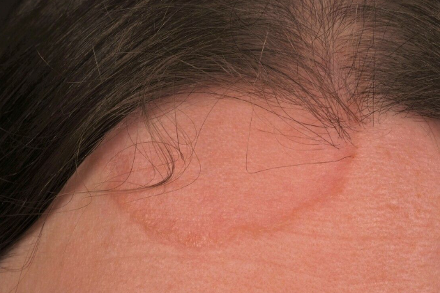
Actinic granuloma (also known as annular elastolytic giant cell granuloma, O'Brien granuloma) is a benign granulomatous elastolytic disorder of the skin that occurs on sun-exposed areas of the body. It is classified under the ICD-10 code L57.5.
Actinic granuloma occurs predominantly in middle-aged women (from the age of 30, but usually over the age of 40) and less frequently in men. The pathogenesis is not completely understood. Ultraviolet radiation is considered to be the main risk factor for this disease. It is believed that the antigenicity of elastic fibers is enhanced by a specific trigger, such as actinic damage, leading to an abnormal cellular immune response targeting the elastic fibers. Cases of the disease have been reported in patients with diabetes mellitus, necrobiosis lipoidica, sarcoidosis, and rarely in patients with lymphoma or leukemia.The condition manifests as annular plaques with an erythematous peripheral rim measuring 3-5 mm and a slightly atrophic hypopigmented center. It is localized on areas exposed to chronic sunlight exposure: the neck, face, upper trunk, forearms, dorsal aspects of the hands, and less commonly the shins and dorsal surfaces of the feet.
The development of the plaques is preceded by the appearance of individual or grouped red papules, which later merge into annular shapes. The size of the lesions ranges from 1 to 10 cm, occasionally larger. The number of plaques is usually less than 10, with solitary lesions being more common. The condition typically progresses without symptoms, although mild pruritus may be observed in rare cases.
Lesions gradually increase in size over several months to years, often followed by spontaneous remission with patchy hypopigmentation or normal skin. Relapses have been reported.
There are clinical variants:- Typical: The most common variant characterized by annular plaques on sun-exposed areas of the body. The lesions grow slowly and spread by the appearance of new lesions.
- Papular: Papules instead of annular plaques.
- Granuloma annulare
- Necrobiosis lipoidica
- Sarcoidosis
- Erythema annulare centrifugum (EAC)
- Tinea corporis
- Nummular eczema
- Secondary syphilis
- Subacute cutaneous lupus erythematosus (SCLE)
- Rheumatoid nodule
- Granulomatous dermatitis

