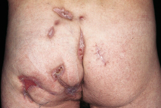
Actinomycosis is an infectious dermatosis that occurs when Actinomycetes bacteria penetrate the skin. It is characterized by chronic suppurative non-contagious lesions of the skin, bones, and internal organs. ICD-10 code: A42.
Actinomycosis is widespread in various geographic regions. Exogenous infection occurs through skin, mucous membranes, bones, and soft tissue injuries. Actinomyces organisms reside continuously in the body of practically healthy individuals, colonizing the oral cavity, tonsillar crypts, bronchi, gastrointestinal tract, and vagina, serving as a source of endogenous infection. There have been no documented cases of actinomycosis transmission from humans or animals.
The causative agents of actinomycosis are gram-positive bacteria, microaerophilic, aerobic, and anaerobic Actinomyces species.
Micro- and macrotrauma, chronic inflammatory processes, presence of stones in ducts, and other factors play a significant role in the development of actinomycosis. For example, in the development of cervicofacial actinomycosis, traumatic factors, post-traumatic tooth extraction, periapical granulomas, presence of salivary stones, and anatomical anomalies (such as branchiogenic neck fistulas) are of great importance. Thoracic actinomycosis is preceded by chest trauma and surgical interventions, while abdominal actinomycosis is associated with appendectomy, cholelithiasis, wounds, contusions, enterocolitis, fecal stones, and others. In 5% of cases, inflammation of the appendix is caused by saprophytic Actinomyces. The development of genital actinomycosis is associated with long-term use of intrauterine devices, which act both as a traumatic factor and sometimes as a carrier of infection. Actinomycosis of the urinary system is usually caused by the presence of stones in the urinary tract and chronic inflammatory diseases. Pararectal actinomycosis is closely related to the condition of the rectum, the presence of epithelial-coccygeal cysts, chronic purulent hidradenitis of the groin and perineal areas, hemorrhoidal nodes, and anal fissures. The disease is characterized by a prolonged course, formation of specific granulomas with subsequent abscess formation and the formation of fistulae with purulent discharge in soft and bony tissues. Secondary bacterial flora, exacerbating the course of the disease and leading to impaired organ function, anemia, intoxication, and amyloidosis, is present in 70-80% of cases along with the actinomyces bacteria.Actinomycosis constitutes 5-10% of chronic suppurative diseases. Among all cases of actinomycosis, 20% involve visceral lesions, while approximately 80% affect the face and neck. Regardless of the localization, actinomycosis is characterized by a gradual development of a dense, sometimes board-like infiltrate without clear boundaries, followed by abscess formation and the formation of one or several fistulas. The discharge from the fistulas is purulent and bloody, odorless, and sometimes granules (grains) of yellow or white color up to 2-3 mm in diameter can be seen. The fistulous tracts are branched (well visualized by fistulography), tortuous, filled with pus and granules. Pain syndrome is minimal. The orifices of the fistulas are retracted or elevated above the level of the skin. Without treatment, the process spreads to the surrounding tissues.
In the neck, axillary and groin areas, fold-like wrinkles are formed and the skin color becomes purplish-blue. The addition of secondary bacterial infection exacerbates the process with a characteristic clinical picture of inflammation.
The human appendix has been shown to contain actinomyces, which under certain conditions cause a disease that in its early stages is mistaken for an appendicular infiltrate. Sometimes, actinomycosis is diagnosed only after fistulae are found on the anterior abdominal wall and the discharge from the fistulae is examined.
Chronic purulent hidradenitis of the axilla and groin is often complicated by actinomycosis and associated with breast involvement. Isolated actinomycosis of the breast also occurs.
Around the pathogen in the sacrococcygeal area, under favorable conditions, there is a slow formation of a specific granuloma with multiple microabscesses and the formation of characteristic tortuous fistulous tracts, which may extend to the pararectal area and rectum.
Rare forms of the disease include actinomycosis of the middle ear, mastoid process, auricle, tonsils, nose, pterygopalatine fossa, thyroid, orbit and eye membranes, tongue, and salivary glands. Involvement of the brain and spinal cord, pericardium, liver, and other areas has been observed. Despite the variety of localizations, an actinomycotic focus follows general developmental patterns characterized by a sequential progression of stages during the course of the disease: infiltrative stage, abscess formation stage, fistulous stage, which in turn leads to an even greater variety of clinical manifestations of actinomycosis.- Actinomycosis of the maxillofacial area and skin is differentiated from abscess, non-specific and tuberculous lymphadenitis, osteomyelitis of the jaws, osteoblastoma, chronic atheromatosis, vegetative pyoderma gangrenosum, conglobate acne, and others.
- Thoracic actinomycosis can present as catarrhal or suppurative bronchitis, pleuropneumonia, lung abscess, encapsulated pleurisy, rib osteomyelitis, and sometimes mimic lung neoplasm. This form of actinomycosis is also differentiated from tuberculosis, aspergillosis, nocardiosis, histoplasmosis, and other diseases.
- Differential diagnosis of actinomycotic breast involvement should be performed with mastopathy, purulent mastitis, abscess, and tumor.
- Abdominal actinomycosis should be distinguished from appendiceal infiltrate, postoperative ligature sinus, interintestinal abscess, peritonitis, Crohn's disease, liver abscess, tumor, and others.
- Pararectal actinomycosis is differentiated from epithelial-coccygeal cyst, buttock abscess, paraproctitis, suppurative atheroma, furunculosis, pararectal sinuses, skin tuberculosis, blastomatous processes.
- Genital actinomycosis should be distinguished from non-specific inflammatory processes, retroperitoneal cellulitis, tuberculosis, chronic salpingitis, uterine fibroids, tubo-ovarian tumor, uterine and adnexal cancer, ectopic pregnancy, as well as from appendiceal infiltrate, pyosalpinx, furunculosis, vaginal and rectovaginal sinuses, chronic pyoderma of external genitalia, Bartholin's abscess, and others.
- Anti-inflammatory treatment with antibiotics based on the sensitivity of the flora. Penicillin-based drugs are used less frequently due to resistance. Preference is given to cephalosporins, tetracyclines, and aminoglycosides in age-appropriate doses during exacerbation (abscess formation) for courses of 2-3 weeks.
- Treatment of concomitant diseases.
- Surgical treatment. Radical and palliative surgeries are performed only after the control of inflammatory processes in the focus of actinomycosis. In the postoperative period blood transfusions and physiotherapy procedures are performed as indicated, and dressings are changed daily. Sutures are removed on the 8th-10th day. The prognosis is more favorable if treatment is initiated in the early stages of the disease with adequate immunotherapy using actinolysate.
- Maintain oral hygiene, avoid putting foreign objects in the mouth that may potentially contain actinomyces (such as straws, toothpicks, etc.).
- Avoid skin and mucous membrane injuries (the entry points for infection).
- Seek timely medical treatment for chronic inflammatory skin conditions, in the oral cavity, thoracic and abdominal areas, genital and pararectal regions.
- Use appropriate prophylactic antibiotic therapy in cases of traumatic and complicated tooth extractions, fractures, appendectomies, surgeries in the pararectal and sacrococcygeal areas, and other surgical interventions.
- Limit the duration of continuous intrauterine device (IUD) use.

