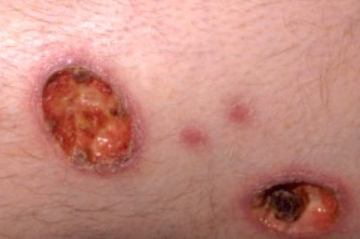
Cutaneous amebiasis is an infection caused by the dysentery amoeba Entamoeba histolytica, resulting from its presence on the skin through the gastrointestinal tract, hematogenous route, or sexual contact. The ICD-10 code for this condition is A06.7.
Approximately 50 million people worldwide are affected by intestinal amebiasis every day. The highest incidence is found in Central and South America, Africa, the Middle East and India. The disease is more common in rural areas and regions with poor sanitation.
Skin involvement occurs in less than 1% of patients with amebiasis. The skin can be affected in the following cases:
Secondary infection:- In most cases, skin lesions in amebiasis occur due to the penetration of dysentery amoebae, which are abundant in the patient's feces, into macerated skin.
- Hematogenous spread: Amoebae can enter the skin hematogenously during surgeries involving the large intestine, hepatic amebiasis, empyema, and pericardial effusion.
- Skin infection can occur through sexual contact, including anal sex and oral-anal contact.
Primary skin infection by direct inoculation is rare. There have been isolated cases of primary cutaneous amebiasis, including on the face.
Sexually active men who have sex with men and persons with HIV/AIDS are at increased risk of transmission. Cutaneous complications are more common in immunocompromised individuals.Common symptoms of amebic colitis occur 7-21 days after infection and include diarrhea, abdominal pain, fever, weight loss, and tenesmus. Skin lesions in secondary infection manifest as the appearance of singular erosions or deep, painful ulcers of various sizes and shapes with sharply elevated undermined edges in the buttocks, groin, and perianal area. Initially, their base is clean, then it becomes covered with serosanguinous or hemorrhagic-purulent exudate with a foul odor. Sometimes, a whitish-necrotic or rarely brownish-necrotic crust forms in the center of the ulcers. Ulcers may communicate with each other through fistulae. Extensive tissue destruction is an important feature of these ulcers. Amebic ulcers have a chronic course with no tendency to heal spontaneously. In rarer cases, cutaneous amebiasis may present as wart-like lesions.
Penile involvement begins as a solitary vesicle, typically on the foreskin, which soon ulcerates, forming a painful ulcer with undermined edges and a central thick crust with purulent discharge.- Pyoderma gangrenosum
- Deep mycoses
- Chancre
- Squamous cell carcinoma
- Genital herpes
- Donovanosis
- Primary syphilis
- Candidiasis
- Crohn's disease
- Ulcerative colitis
- Behçet's disease
- Extramammary Paget's disease
- Metronidazole: 500-750 mg three times a day for 7-10 days. For children, the dosage is 35-50 mg/kg/day divided into three doses for 10 days.
- Tinidazole: 2 g per day for 3-6 days.
- Ornidazole: 500 mg twice a day for 5-10 days.
- Intramuscular injection of a 2% solution of emetine hydrochloride: 1.5-2.0 ml twice a day for 7 days (2-4 courses with a 7-10 day interval).
- Diiodohydroxyquinoline: 650 mg three times a day after meals for 20 days.
- Paromomycin: 0.5 g two to three times a day for 5-7 days. For children, the dosage is 10 mg/kg two to three times a day for 5-7 days.

