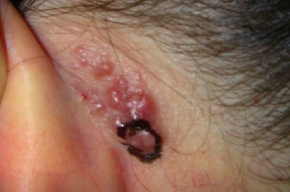
Angiolymphoid hyperplasia with eosinophilia (ALHE) is a benign skin disorder of unknown etiology. It is characterized by the development of papules and nodules due to lymphoid infiltration and unusual benign angiomatous proliferation. The ICD-10 code for ALHE is D18.0.
Previously, it was considered a variant of Kimura's disease, but it is now recognized as a distinct entity. The exact cause of ALHE is unclear, and it is thought to have a reactive or neoplastic nature. It primarily affects adults, with a higher incidence in women between the ages of 20 and 30. Some patients report a history of previous skin trauma. Vascular malformations and arteriovenous shunts are thought to play a role in the pathogenesis of the disease.The disease is characterized by the appearance of papules or nodules of pink, dull red, brown and brownish color with a smooth surface in the head and neck area, especially around the earlobes and on the scalp. Less commonly, the eruptions are found on the trunk, mouth (lips, gums, palate, tongue), distal parts of the limbs, skin folds, vulva, and penis.
Dermal papules have a semi-spherical shape, clear borders, firm elastic consistency, and in rare cases there may be a central umbilication. Palpation of the papules may sometimes cause slight tenderness. Occasionally the papules may coalesce into plaques. Multiple eruptions occur in approximately more than half of cases. Subcutaneous nodules are located under the skin and may be large, up to 5-7 cm, with less pronounced pigmentation compared to papules.
Patients complain of itching, burning, and throbbing pain in the affected areas, but the disease is often asymptomatic. Regional lymphadenitis is seen in some cases. Blood eosinophilia occurs in 10-15% of cases. Extracutaneous involvement in the form of areas of osteolysis and osteosclerosis is most commonly seen in long and small tubular bones and the spine. Cases of extracutaneous localization of the disease have been described in the breast, testes, ovaries, lymphatic vessels, and arteries.The diagnosis is made based on the clinical presentation, medical history, and histological examination, which reveals a benign angiomatous or angiomatoid proliferation within a stroma that is extensively infiltrated by lymphocytes and eosinophils. The infiltrate contains lymphoid follicles with prominent germinal centers. Multiple arteries and veins are observed, lined with enlarged edematous, vacuolated endothelial cells with large epithelioid and histiocytoid nuclei, along with intravascular proliferation of endothelial cells.
Blood testing is recommended to evaluate for eosinophilia and IgE levels. In rare cases, radiography of the tubular bones may be necessary.- Kimura's disease
- Sarcoidosis
- Cylindroma
- Scleromyxedema
- Leiomyoma
- Eosinophilic granuloma
- Recurrent (chronic) polychondritis
- Jessner lymphocytic infiltration of the skin (JLIS)
- Angiosarcoma
- Kaposi's sarcoma
- Hypereosinophilic syndrome.

