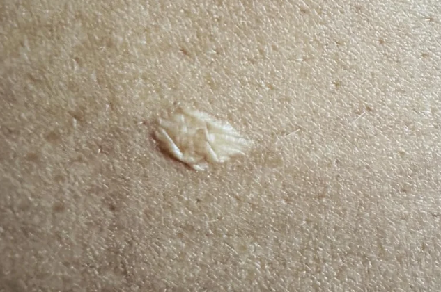
Anetoderma is a distinct form of skin atrophy that develops without apparent cause. It is coded as L90.2 in the ICD-10 classification.
The condition predominantly affects women, especially those in early adulthood, between the ages of 20 and 30. The etiology and pathogenesis of anetoderma are not fully understood. It is believed that the development of the condition is rooted in a constitutional abnormality, possibly related to endocrine disorders which subsequently affect the elastic and collagen tissues as well as the nerves of the skin. Some opinions suggest that anetoderma is a form of idiopathic progressive cutaneous atrophy with small macules.
There is evidence supporting the involvement of immune mechanisms in the development of anetoderma, indicated by the frequent presence of plasma cells in the infiltrates, abundant T-cells with a predominance of T-helper cells, and features of leukoclastic vasculitis with perivascular deposition of IgG, IgM, and complement components. Considering the occurrence of macular atrophy in various conditions such as urticaria pigmentosa, xanthomas, long-term corticosteroid therapy, penicillamine use, and regression of many dermatoses (tuberculosis, leprosy, etc.), it can be hypothesized that anetoderma represents a heterogeneous condition, with the destruction of elastic fibers as a common underlying process influenced by various causes. Moreover, several studies indicate the existence of more widespread forms of anetoderma involving other organs, not limited to skin involvement alone.Traditionally, several clinical variants are distinguished:
- Atrophic patches that develop after a preceding erythematous stage (classical Jadassohn type).
- Atrophic lesions occurring at the site of urticarial or edematous elements (Pellizzari type).
- Atrophic patches on clinically unaffected skin (Schwenninger-Buzzi type).
However, this classification does not seem to be sufficiently justified. The inflammatory response may be so mild that patients are unaware of the disease until the atrophic stage, when herniated protrusions appear. Different variants may coexist in the same patient. Atrophic macules can occur on any part of the skin, but are more common on the upper trunk, arms, and face. The atrophic macules are small, typically 1-2 cm in diameter, with round or oval borders, a whitish-blueish color, and a shiny, wrinkled surface. A characteristic feature is the hernia-like protrusion, and when pressure is applied with a finger in this area, it gives the impression of a hollow or empty space. However, this feature is not present in all lesions; some may show a depression instead. The number of atrophic patches varies from individual to individual, from a single lesion to dozens. Subjective disturbance is absent.
The classic type of Jadassohn's anetoderma is characterized by the appearance of single or multiple purplish-pink or yellowish-pink patches with irregular round borders, measuring up to 1-2 cm in diameter. These lesions are more common on the trunk, upper extremities, and face. Gradually, without subjective sensation, atrophy develops in the center of the patch. The skin in these areas becomes pale and wrinkled, resembling crumpled cigarette paper, and the lesion protrudes slightly above the level of the surrounding skin. Over time, white, shiny, scar-like patches remain at the sites of the primary lesions, which may evolve into soft, hernia-like protrusions of the skin that give a sensation of hollowness to palpation (the finger easily sinks into the lesion).
Schwenninger-Buzzi type anetoderma is clinically characterized by hernia-like protruding atrophic patches, mainly on the back and upper extremities. The "tumor-like" appearance and bluish-white color of the atrophic lesions are emphasized. In contrast to the classic Jadassohn type of anetoderma, the atrophic patches in this type protrude significantly beyond the surrounding skin. Telangiectasias may be present on the surface of the lesions, and the initial inflammatory phase is always absent.
Pellizzari's urticarial type is rare, and anetoderma develops at the site of wheals. The eruptions, usually without subjective sensations, form sac-like protrusions, and when palpated, the finger seems to sink into an empty space, leaving the surface of the patch slightly depressed for some time.
Anetoderma has a chronic course with periodic appearance of new lesions, while old ones retain their activity even after more than 15 years of existence. Anetoderma is a component of Blegvad-Haxthausen syndrome, which includes atrophic macules, blue sclerae, bone fragility, and cataracts. There have been reports of its association with bilateral congenital nephropathy in a 2-year-old child with a fatal outcome. Patients should undergo further evaluation to exclude associated diseases of the eyes, bones, heart, lungs, gastrointestinal tract, and endocrine glands, as well as systemic lupus erythematosus. However, there is currently insufficient evidence to establish a non-random association between these conditions.The diagnosis is based on the presence of small round or oval areas of skin atrophy with characteristic herniated protrusions.
Histopathology reveals a decrease in the amount, fragmentation, or absence of elastic fibers. In the early stages, there may be mild perivascular infiltrates, predominantly lymphocytic, with some granulocytes present.- Atrophy. The diagnosis of secondary scar atrophy is based on the patient's history, the characteristics of scar changes, and the presence of typical manifestations of the underlying dermatosis that led to scar formation.
- In dystrophic epidermolysis bullosa, white nodular elements with a diameter of 1-1.5 cm and a wrinkled surface may be observed; however, they are firm and show a follicular pattern in their area.
- Morphea. In morphea, the plaques are usually large in size, and atrophic changes are only detected in the late stage of the disease. In the early stages, there is thickening of the plaques, and the presence of a livid ring at the periphery is characteristic. Histologically, the main changes are observed in collagen fibers rather than elastic fibers.
- Neurofibromatosis. In neurofibromatosis, in addition to herniated protrusions overlying neurofibromas, there are other signs of the disease such as pigmented spots, fibromas, etc.
- Atrophoderma of Pasini-Pierini. In atrophoderma of Pasini-Pierini, the spots are few in number, mainly located on the back, and typically have large sizes (up to 10 cm or more). There is no herniated protrusion.
There are no effective treatments for anetoderma.
In the early stages, when erythema is present, penicillin or tetracycline may be prescribed at a dose of 1,000,000 units per day for 10-14 days. For active progressive forms of anetoderma, general tonic and vitamins A and E are recommended. (Not evidence based. Need more investigations)
There is evidence of good therapeutic results with the use of aminocaproic acid in the treatment of anetoderma, and cessation of disease progression has been observed after a single administration of corticosteroids.
