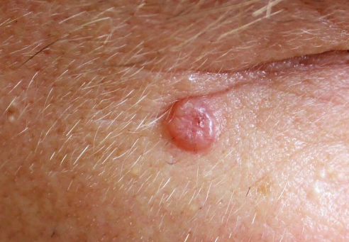
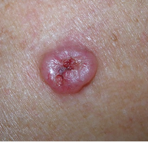
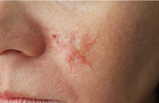
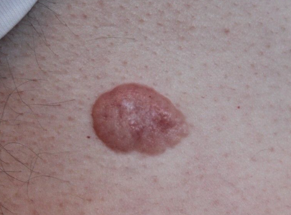
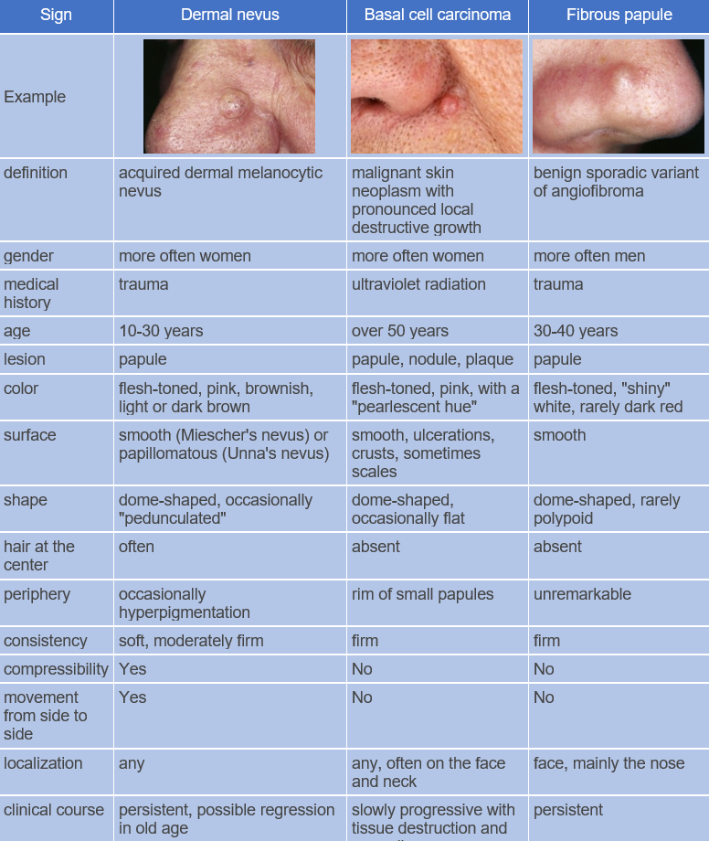
Basal cell carcinoma is a common skin tumor with pronounced local destructive growth, a tendency to recur, but generally no metastasis. It is localized to exposed areas of the skin, such as the face, neck, and scalp. The ICD-10 code for this condition is C44.9.
The disease is more common in people over the age of 50, but it can also develop at a younger age. It occurs with equal frequency in both women and men. The histogenesis of the tumor is still debated. Most researchers adhere to the dysontogenetic theory of origin, according to which basal cell carcinoma develops from multipotent epithelial cells. These cells can differentiate and form various structures. The development of basal cell carcinoma is also influenced by genetic factors, immune disorders, and adverse environmental exposures (such as intense sunlight and exposure to carcinogenic substances). The tumor can develop on visually unaltered skin as well as in the presence of various skin conditions (actinic keratosis, post-radiation dermatitis, lupus vulgaris, nevi, psoriasis, etc.).
Basal cell carcinoma can be classified into:- Pigmented
- Nodular
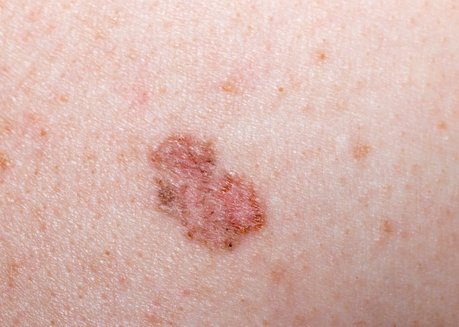
- Superficial
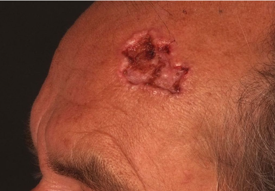
- Ulcerative (ulcus terebrans): BCC can have destructive growth, penetrating deeper layers of the skin.
- Sclerodermiform (morphoeic)
- Fibroepithelial: BCC can have fibrous and epithelial components.
Nodular BCC
Ulcerative BCC
Superficial BCC
Sclerodermiform BCC
Fibroepithelial BCC
- Nodular BCC: Benign tumors of sweat glands and hair follicles.
- Ulcerative BCC: Squamous cell carcinoma, leishmaniasis.
- Pigmented BCC: Lentigo and lentigo maligna.
- Superficial BCC: Bowen's disease, chronic focal inflammatory dermatoses, actinic keratosis, discoid lupus erythematosus.
- Sclerodermoid BCC: Scarring skin lesions.
- Fibroepithelial BCC: Fibroepithelial polyp, solid trichoepithelioma, dermatofibroma.
Surgical excision, Mohs surgery, interferon therapy, local chemotherapy
Prognosis
Overall, the prognosis is favorable since BCC rarely metastasizes unless it transforms into a metatypical form. The recurrence rate is quite high, with some reports indicating rates of up to 17-20%.
