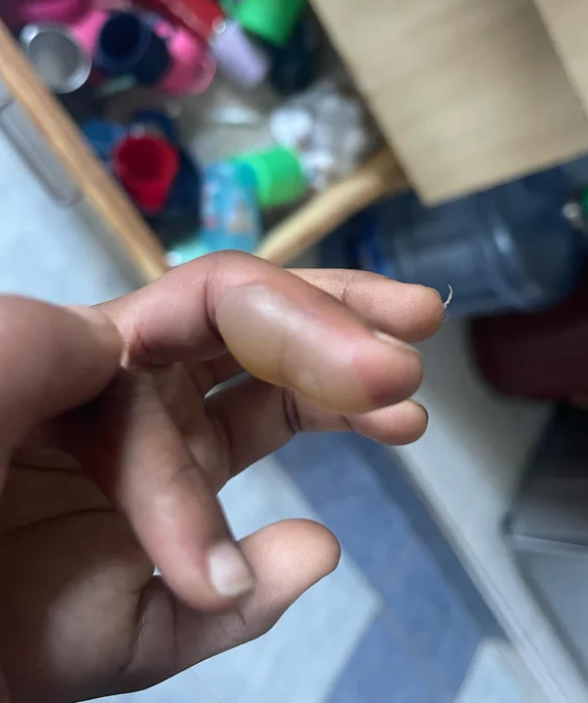
Blistering distal dactylitis is a localized bacterial skin infection characterized by the formation of superficial blisters on the distal surfaces of the fingers and, less commonly, toes. It is primarily caused by Group A beta-hemolytic Streptococcus and is coded as L08.9 in ICD-10.
The condition is commonly seen in children between the ages of two and 16, although cases have been reported in infants and adults. The infection is typically caused by beta-hemolytic group A streptococcus, less commonly by group B streptococcus or Staphylococcus aureus (including methicillin-resistant strains).
The portal of entry for infection is usually skin damage resulting from insect bites, trauma, erosions, or burns. It is thought that infection may also occur by autoinoculation during nose picking, as the nasal sinuses may serve as a reservoir for staphylococcal infection, or by finger sucking in cases of streptococcal pharyngitis.
The exact pathogenesis of blistering is unclear. While the production of exfoliative toxins (exfoliatin) is involved in the formation of blisters in staphylococcal infections, streptococci do not appear to cause blistering by toxins or other mechanisms. Some authors consider bullous dactylitis to be a variant of bullous impetigo. Immunodeficiency and diabetes mellitus are common in adult patients with this condition.The condition is characterized by the appearance of superficial, solitary, or occasionally multiple blisters with a firm base. They are oval in shape, 10 to 30 mm in size and filled with seropurulent fluid. Blister formation is preceded by redness of the skin and is followed by an erythematous halo.
The typical location is the palmar surface of the distal phalanx of the finger, particularly in the area of the finger pad, commonly affecting the index finger and less frequently the toes. Over time, the condition progresses with the enlargement of the blister to involve the lateral surfaces of the finger and the periungual folds, leading to its rupture and the formation of erosions. Rarely, the process may extend to the palms, dorsum of the hand, soles, or dorsum of the foot. Pain and systemic symptoms are usually absent.
The disease has a benign course, although isolated cases of self-amputation of the finger have been reported. Recurrence of the disease is possible in individuals with chronic sites of infection or carriers of Staphylococcus and Streptococcus bacteria.- Herpetic infection (herpetic whitlow)
- Deeply located clustered blisters surrounded by an erythema zone, characterized by pain
- Possible history of recurrent eruptions in the same location
- PCR reveals herpes simplex virus
- Regional lymph nodes may be affected
- Acute paronychia
- Erythema and swelling in the lateral and proximal nail folds (no prominent blister)
- The affected area is usually painful
- Bacteria culture test reveals Staphylococcus aureus
- • Hand-foot-mouth disease
- Characteristic rash localized on the lateral surfaces of the fingers or palms and soles
- Blisters have more oval contours and are deeper
- Multiple and smaller blisters are present
- Often associated with oral erosions
- Bullous epidermolysis
- Recurrent blister formation triggered by trauma In the Weber-Cockayne type, eruptions predominantly occur on the palms and soles, but widespread involvement is typically characteristic (with multiple eruptions)
- Other types of bullous epidermolysis have additional rash locations on the body or mucous membranes
- Burn
- Bullous impetigo
- Superficial blisters
- Blisters from finger-sucking or shoe friction (on the feet)
- No signs of inflammation
- The content of the blisters is serous (clear)
- Erysipeloid
- Orf
Topical therapy:
- Incision or puncture of blisters followed by application of moist dressings with antiseptic and antibiotic solutions.
Systemic therapy:
In case of unknown pathogen:
- Cephalexin 25-50 mg/kg/day orally every 6 hours, with a maximum daily dose of 4 g for 10 days.
In case of detection of Staphylococcus aureus:
- Amoxicillin/clavulanic acid 20-40 mg/kg/day orally every 8 hours for 10 days.
In case of detection of Group A Streptococcus:
- Penicillin V 25 mg/kg/day every 6-8 hours, with a maximum daily dose of 3 g for 10 days.
- Cephalexin 25-50 mg/kg/day orally every 6 hours, with a maximum daily dose of 4 g for 10 days.
- Erythromycin 30-50 mg/kg/day orally every 6 hours, with a maximum daily dose of 2 g for 10 days.
- Clindamycin 15 mg/kg/day orally every 6 hours for 10 days.

