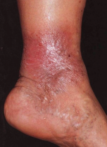
Stasis dermatitis is a form of eczema affecting the lower extremities, associated with edema, varicose and dilated veins, and hyperpigmentation. It is classified under ICD-10 code I83.1.
A history of deep vein thrombosis, surgical trauma or ulcer formation is common, as is exposure to irritants or potential allergens in the wound care products used by the patient. There is often a family or personal history of varicose veins.
Predisposing and aggravating factors include obesity, diabetes mellitus, congestive heart failure, previous leg fractures, venous hypertension, congenital absence of venous valves, anemia and smoking.Patients complain of heaviness and pain in the legs that worsens with walking and prolonged standing, with edema toward the end of the day. On examination, the veins of the affected limbs appear dilated and tortuous when standing. Dermatitis and pruritus are characteristic.
Stasis dermatitis occurs on the shins or around venous ulcers. The dermatitis is well defined and associated with dry, cracked, erythematous skin. Typical clinical signs include edema, purpura, brown pigmentation (hemosiderosis), painful erosions or ulcers with clear margins. Skin involvement is usually bilateral but may be unilateral. Patients suffer from severe pruritus and excoriations may lead to secondary infections.
Stasis dermatitis is a chronic condition that usually recurs, especially after irrational topical therapy and water exposure.
Complications and atypical course: Dermatitis may become generalized (id reaction) if the condition persists. Small areas of atrophy and depigmentation may appear as star-shaped atrophy (white atrophy of Blanche). White scars in the medial calf area indicate previous ulceration. Stasis papillomatosis (elephantiasis nostra) and verrucous hyperplasia are seen in cases of chronic stasis in the limbs due to local lymphatic disorders such as chronic venous insufficiency, primary lymphedema (Milroy's disease), trauma, and chronic scarring inflammation. Secondary Staphylococcus aureus infection is typical, especially on excoriated skin. As the condition progresses, lipodermatosclerosis may develop due to chronic ischemia of adipose tissue.Diagnosis is based on the clinical picture and medical history.
Indicated tests include:
- Duplex ultrasound with color mapping.
- Measurement of the brachial index to detect arterial insufficiency; a result of less than 0.8 indicates significant arterial insufficiency.
- Coagulation tests - for patients with a history of severe multiple deep vein thrombosis.
- Histological examination.
- Cellulitis (key features: sudden pain and severe swelling).
- Xerosis (dry skin).
- Contact dermatitis - especially to neomycin, bacitracin, fragrances, and preservatives found in popular moisturizing lotions and hydrocortisone; allergic contact dermatitis is typically associated with stasis dermatitis.
- Erythema nodosum.
- Asteatotic eczema.
- Atopic dermatitis.
- Fungal infection (KOH test for fungi).
- Granuloma annulare.
- Necrobiosis lipoidica.
- Pretibial myxedema.
- Nummular eczema.
- Psoriasis.
- Lichen simplex chronicus
- Capillaritis and other pigmented purpuric dermatosis.
- Vasculitis.
- Lymphedema.
- Hypertrophic lichen planus.
- Cool water compresses applied for 10-20 minutes twice a day during acute exudative inflammation.
- Topical steroids are effective for dermatitis or inflammatory components. They are applied twice a day (cream or oil for acute inflammation, ointment for chronic inflammation) for 2-3 weeks.
- Aseptic dressings, topical antibiotics (mupirocin), and a course of systemic antibiotics (doxycycline, cephalexin) are prescribed for infection (impetiginization).
- Systemic steroids are prescribed for 3 weeks with a gradual dose reduction in cases of generalized dermatitis (id reactions).
- Systemic antihistamines 10-25 mg every 4-6 hours can be used for intense itching.
- Use of emollients helps combat dry skin. Vaseline-based emollients such as petroleum jelly, Aquaphor, Aveeno, and Cetaphil cream are preferred over lotions. Popular moisturizers may contain fragrances and preservatives that can aggravate contact allergies.
- Special compression stockings or tight bandages (20-30 mmHg) are used. In some cases, higher compression (30-40 mmHg on each calf) may be required. If the patient is not mobile enough or not strong enough to wear elastic stockings, elastic bandages like "Ace" bandage can be used to reduce edema. They are applied in the morning after getting out of bed.
- Superficial venous insufficiency may decrease after subcutaneous vein removal or sclerotherapy.

