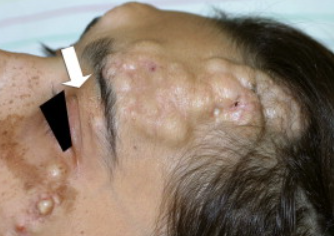
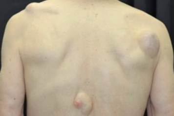
Pilar cyst (trichilemmal cyst) is a benign developmental anomaly of the hair follicle. ICD-10 code: L72.1.
It may be present from birth, but more commonly appears in old age, sometimes reaching significant sizes. It occurs in 5-10% of population, predominantly in middle-aged women. Familial forms are often observed and are probably inherited in an autosomal dominant pattern.
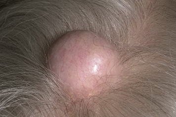
Familial cases with autosomal dominant inheritance have been reported. A mutation in the TRICY1 gene on chromosome 3q24-q21.2 is presumed. Associations with glomangioma, melanocytic nevus, and Jadassohn sebaceous nevus have been described.Single pilar cyst
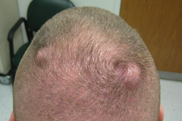
Multiple pilar cysts
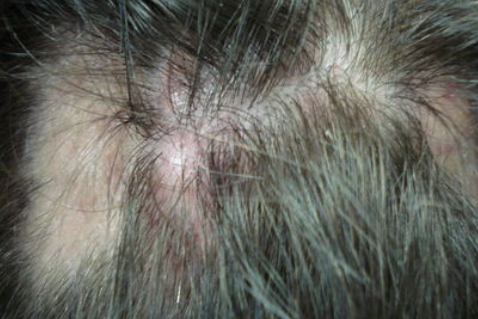
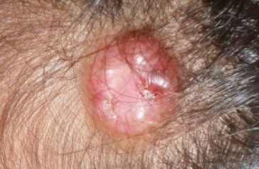
Proliferating pilar cyst
Proliferating pilar cyst, also known as proliferating trichilemmal tumor, is a rare neoplasm that develops from a pre-existing trichilemmal cyst after trauma or inflammation and arises from hair matrix cells. It can also occur de novo (on unaltered skin) and in the setting of nevus sebaceous.
Clinically, it presents as a single soft, smooth-surfaced, flesh-colored nodule or plaque. Multiple lesions are seen in approximately 10% of cases. The size ranges from 1 to 25 cm (average 5 cm). In 90% of cases, it is located on the hairy part of the scalp, less commonly on the face, trunk, upper limbs, vulva, or buttocks.
The course is unpredictable and the process may be benign and slow or very rapid with histologic signs of malignancy (malignant proliferating trichilemmal tumor) with metastases to cervical and supraclavicular lymph nodes and soft tissues of the neck.
Tricholemmocystic nevus
Tricholemmocystic nevus (nevus tricholemmocysticus) is an organoid epidermal nevus characterized by the presence of tricholemmal cysts in the form of yellowish or flesh-colored plaques along Blaschko's lines, along with multiple small verrucous hyperkeratotic papules. Lesions are found on the head, neck, trunk, extremities, palms, and soles. When localized on the scalp, diffuse alopecia is observed.
It is usually identified at birth, occasionally in early childhood. The etiopathogenesis is not well understood.
FLOTCH syndrome
- Lipoma
- Fibrolipoma
- Epidermal cysts
- Trichoepithelioma
- Multiple steatocystoma
Surgical excision or electroexcision with mandatory removal of the capsule.
Prognosis is favorable.

