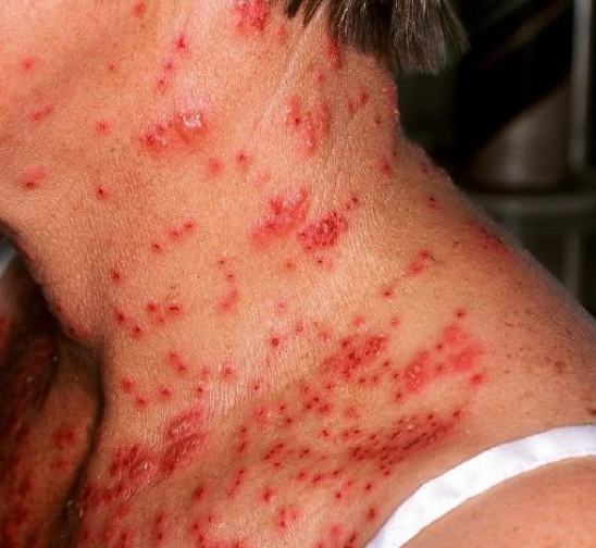
Eczema herpeticum Kaposi (Kaposi's varicelliform eruption) is a viral skin infection characterized by a widespread vesicular rash localized in areas of pre-existing dermatoses. It is coded as B00.0 in ICD-10.
The disease occurs in individuals from 5 months to 70 years of age, but is more common in children and young adults up to 30 years of age (with a median age of 15-24 years), with no gender or ethnic differences. The disease is caused by infection with herpes simplex viruses 1 and 2, varicella-zoster virus, Coxsackie A6 and A16 (Coxsackie eczema), and cowpox virus (vaccinia eczema). Infection occurs through contact with carriers and individuals with clinical manifestations of these infections. The prevalence ranges from 4.03 to 7.30 per one million hospitalizations.
The exact pathogenesis is not fully understood. However, it is well established that herpetic eczema develops in patients with the following dermatoses:
Commonly Associated Dermatoses:
- Atopic eczema
- Darier disease
- Hailey-Hailey disease
- Pemphigus foliaceus
Rarely Associated Dermatoses:
- Seborrheic dermatitis
- Rosacea
- Acne
- Ichthyosis
- Congenital ichthyosiform erythroderma
- Grover disease
- Allergic contact dermatitis
- Irritant contact dermatitis
- Psoriasis
- Dowling-Degos disease
- Cutaneous T-cell lymphoma
- Bullous pemphigoid
- Wiskott-Aldrich syndrome
- Sézary syndrome
- Burns and injuries
- Staphylococcal scalded skin syndrome
- Cosmetic procedures (dermabrasion and laser therapy)
It is believed that the weakening of the skin barrier function due to dermatoses and impairment of the immune system (cellular and humoral) are the main components of the disease's pathogenesis.
Risk Factors:
- Early onset of atopic dermatitis
- Localization of dermatoses on the head and neck
- Large surface area of dermatosis lesions
- High levels of IgE
- Colonization of Malassezia sp.
- Low levels of NK cells and IL-2 receptors
- Low levels of antimicrobial peptide cathelicidin LL-37, human β-defensin, and gamma-interferon in the skin
- Mutations in IL-18 and IL-10 genes
- Prolonged use of calcineurin inhibitors (pimecrolimus and tacrolimus), cyclosporine, methotrexate, and retinoids.
After an incubation period of 5-19 days (average 10 days), sudden eruptions occur in areas of pre-existing dermatosis or injury. The eruptions appear as scattered, disorderly vesicular rashes with a central umbilication on an edematous and erythematous background. After 2-3 days, some of the vesicles, filled with hemorrhagic content, rupture, forming sharply painful "punched-out" small moist erosions that quickly become crusted, often with hemorrhagic crusts. When erosions merge, larger poly-cyclic lesions are formed. About 5-7 days after the onset of the disease, new eruptions appear, spreading beyond the initial areas of dermatosis, and in most cases the process takes on a generalized character.
Fever up to 39-40°C is characteristic and usually appears on the 2nd or 3rd day of the disease, accompanied by general symptoms such as malaise, weakness and headache, which last for 4-7 days. Pain, burning, or itching may be observed at the site of the eruption. Regional lymphadenitis is common. After 14-18 days, sometimes longer, the crusts dry and fall off, leaving pink or hyperpigmented patches and, extremely rarely, superficial scarring. The typical duration of the primary episode of herpetic eczema is 2-6 weeks (average 16 days).
In some cases, the disease may be recurrent, with episodes occurring with varying frequency, but these episodes are usually less severe. A seasonal pattern has not been observed.
Complications of herpeticum eczema include herpes labialis involving the mucous membranes of the mouth, pharynx, larynx, trachea, liver, lungs, gastrointestinal tract, and meninges. In severe cases, adrenal necrosis and shock may occur.
Secondary infection most commonly occurs with Staphylococcus aureus, group A beta-hemolytic Streptococcus, Pseudomonas aeruginosa, and Peptostreptococcus. This results in the appearance of pustular eruptions and, in severe cases, the development of bacterial sepsis, meningitis, encephalitis, pneumonia, and otitis.
Ocular involvement includes blepharitis, conjunctivitis, keratitis, and uveitis. Herpetic keratitis can lead to blindness due to scarring.
The use of intravenous acyclovir in addition to systemic/local antibiotic treatment has reduced mortality from 50% to less than 10%.- Tzanck smear from the lesion material.
- Identification of viral antigens in vesicular swabs.
- Histological examination
- Determination of type-specific IgM and IgG antibodies in the blood using from the 10th day of the disease.
- Polymerase chain reaction (PCR) - detection of viral DNA from lesion material (in cases of viremia in the blood serum).
- In case of suspected secondary infection and sepsis, bacterial culture with sensitivity to antibiotics is performed.
- Chickenpox (Varicella)
- Disseminated Herpes Zoster (Shingles)
- Bullous Impetigo
- Molluscum Contagiosum
- Folliculitis (Staphylococcal, Pseudomonas, Candidal Folliculitis)
- Rosacea
- Contact Dermatitis
- Dermatitis Herpetiformis
- Drug erupcion
- Acute Generalized Exanthematous Pustulosis (AGEP)
First line:
- Acyclovir orally 400 mg three times a day for 7 days.
- Valacyclovir orally 500 mg three times a day for 7 days.
- Famciclovir orally 250 mg three times a day for 7 days.
Second line:
- Acyclovir intravenously (10-15 mg/kg/day) for 7 days.
Third line:
- Foscarnet at a dose of 120 mg/kg/day for 7 days.
For secondary infection:
- Erythromycin or dicloxacillin orally.
For frequent recurrences:
- Long-term use of low doses of acyclovir or valacyclovir.

