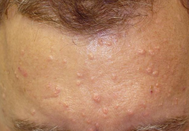
Sebaceous gland hyperplasia is a common benign condition in which the sebaceous glands of the face become significantly enlarged. ICD-10 Code: L73.9
Sebaceous hyperplasia occurs in both men and women. The papules rarely appear before the age of 30, but become more common with age. Approximately 80% of patients over the age of 70 have at least one such lesion.
Most lesions consist of a single hypertrophied sebaceous gland and numerous lobules are arranged around an enlarged sebaceous duct. These lesions can occur on any skin type, but are more prominent on fair skin.
The etiology of sebaceous hyperplasia is unknown, but age-related changes in sebaceous gland function are thought to play a role. Heredity is almost certainly a contributing factor. Sun exposure is thought to be a contributing factor. Occasionally, hyperplasia develops as a result of corticosteroid therapy. The papules can cause facial disfigurement and are primarily a cosmetic concern.The disease begins with the appearance of small flesh-colored or pale-yellow papules, 1-2 mm in size, minimally elevated above the surface of the skin. Over time, they reach a maximum size of 3-4 mm, becoming more distinctly yellowish, with a dome-shaped form and a central umbilication, sharply demarcated from the surrounding skin.
Numerous regularly arranged small telangiectasias can be observed, radiating from the central umbilication in the papules and spreading between them towards their periphery. The central dimple is an almost constant feature. Papules may be solitary but more commonly they are multiple. Yellowish-white papules are typically seen on the forehead, nose, cheeks, and occasionally on the eyelids. The lesions are completely asymptomatic but persist over time.- Basal Cell Carcinoma. In sebaceous hyperplasia, telangiectasias radiate from the lesions in a regular pattern, in contrast to the disorganized arrangement of telangiectasias on the face in basal cell carcinoma. The eruptions in sebaceous hyperplasia have a soft consistency, and when the papules are squeezed from the sides of the central umbilication, a drop of sebum usually appears.
- Milia
- Intradermal nevus
- Flat warts
- Keratoacanthoma
- Syringoma
- Fibrous papules
- Fordyce spots
- Molluscum contagiosum
- Xanthomas
- Xanthelasma
- Pilomatricoma
- Rosacea
- Acne
- Colloid milium
- Trichoepithelioma
No treatment is required, but patients may seek help for cosmetic reasons.
Carbon dioxide laser, shave excision, electrocautery with curettage, cryotherapy, and trichloroacetic acid are effective methods of removal. For successful treatment, the lobules of sebaceous glands located in the superficial layers of the dermis must be destroyed. Excessive treatment can lead to scarring.
Isotretinoin (1 mg/kg/day for 2 weeks), and topical cream with tretinoin 0.025% for 6-18 months can also be used as treatments.
