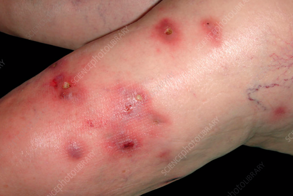
Swimming pool granuloma, also known as fish tank granuloma,aquarium granuloma, is a chronic infectious skin disease caused by Mycobacterium marinum, characterized by the formation of nodules and plaques, mainly on the extremities. Its ICD-10 code is A31.1.
The condition is more common in men under the age of 40, likely associated with certain professions such as fishermen, aquarium keepers, and divers, among others. It is also occasionally found in vacationers on beaches. The estimated prevalence in the United States is 0.27 per 100,000 population.
The disease is caused by Mycobacterium marinum, a non-motile, non-sporeforming, acid-fast bacterium found in marine water, plants, and soil that causes a tuberculosis-like disease in fish. Infection occurs through skin trauma during activities such as swimming in the ocean or swimming pools, handling fish, cleaning aquariums, and contact with reptiles, mollusks, and other marine animals.After a long incubation period (ranging from weeks to months), a papule appears at the site of injury and rapidly enlarges into a reddish-brown nodule or plaque with a warty or scaly surface, 2-4 cm in diameter. Usually one or more lesions are observed, localized on the fingers, dorsum of the hands, forearms, less frequently on the feet and shins, occasionally over joint surfaces (phalangeal, metacarpal, elbow, knee). Rarely, the trunk, face, and neck may be affected.
In most cases, the formation is asymptomatic, occasionally causing itching or tenderness to pressure. Over the course of several months, the lesion breaks down into an ulcer that heals with an atrophic scar.
Less commonly, the disease presents as a sporotrichoid form with multiple nodules linearly aligned along lymphatic vessels, accompanied by lymphangitis, or a disseminated form with fever. The disease has a prolonged course, lasting from several months to several years. In most cases, even without treatment, spontaneous recovery occurs within 3 to 36 months. However, in the sporotrichoid form, the disease can last for decades without treatment.The diagnosis is based on the patient's history, clinical presentation, and results of laboratory tests:
- Tuberculin skin test using purified protein derivative (PPD) shows positive results in 67%-100% of cases.
- Microscopic examination of pus smears stained with Ziehl-Neelsen acid-fast stain detects acid-fast bacilli in 50-60% of cases.
- Polymerase chain reaction (PCR) yields positive results in 90-95% of cases.
- Cultural examination reveals colonies of lemon-yellow color after 14-21 days.
- Histological examination shows a nonspecific pattern - early lesions exhibit inflammatory reactions, lymphocytes, neutrophils, macrophages, hyperkeratosis, and acanthosis, while late lesions display epithelioid cells and Langhans giant cells.
- Sporotrichosis
- Blastomycosis
- Coccidioidomycosis
- Nocardiosis
- Tularemia
- Cutaneous Tuberculosis
- Leishmaniasis
- Actinomycosis
- Sarcoidosis
- Cellulitis
- Mycetoma
- Cutaneous Lymphoma
- Sarcoma
- Bacterial Abscess
- Pyoderma Gangrenosum
- Vasculitis
- Superficial Thrombophlebitis
- Granuloma Annulare
- Hypertrophic Lichen Planus
- Foreign Body Reaction to Sea Urchin Spines, Mollusk Shells
The World Health Organization (WHO) recommends at least 8 weeks of treatment. The average course of treatment is usually 3 months.
Antibiotics are prescribed in the following regimens
- Clarithromycin (500 mg orally twice daily)
- Minocycline or doxycycline (100 mg orally twice daily)
- Rifampicin (600 mg orally once daily) + ethambutol (1.2 g orally)
- Trimethoprim-sulfamethoxazole (500 mg twice daily)
- Levofloxacin (500 mg once daily)

