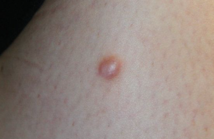
Dermatofibroma (benign fibrous histiocytoma) is a benign skin tumor of connective tissue origin. ICD-10 code: D23.9.
It occurs more frequently in young females, mainly on the lower extremities. Microtraumas and insect bites are considered etiologic factors. The tumor consists of fibroblastic cells, fibrous substances and blood vessels in varying proportions. Fibroblastic elements in the tumor have a phagocytic tendency and often contain lipids and iron.
According to the WHO classification, the following histological variants of the tumor are distinguished:
- Cellular dermatofibroma.
- Aneurysmal dermatofibroma.
- Epithelioid dermatofibroma.
- Atypical (pseudosarcomatous) dermatofibroma.
Dermatofibroma is characterized by solitary, rarely multiple, round nodules up to 1-2 cm in diameter located deep in the skin or subcutaneous tissue. Therefore, the tumor may appear to be embedded in the skin, with its upper pole at the level of the surrounding tissue, or in other cases, it may be domed. The nodules are firm, with a smooth or sometimes verrucous surface, ranging in color from normal skin tone to reddish brown, and they are usually painless. In some cases they may coalesce into lobulated conglomerates.
A characteristic symptom is the "dimple" sign, in which the tumor sinks slightly into the surrounding tissue when pressed between two fingers.
Multiple dermatofibromas are occasionally seen in systemic lupus erythematosus, HIV infection, and may be associated with osteopoikilosis (Buschke-Ollendorff syndrome). An atrophic variant of the tumor has been described, which may mimic atrophia maculosa varioliformis cutis.
Dermatofibromas may persist for many years without spontaneous regression and have a marked tendency to recur locally after excision. However, several cases of histologically proven metastasis to lymph nodes, lungs, and abdominal wall have been documented in the literature, occurring 2-2.5 years after excision of the primary tumor.- Pigmented nevi.
- Infantile systemic hyalinosis.
- Dermatofibrosarcoma protruberans
- Infantile myofibromatosis.
- Melanoma
- Keloid or hypertrophic scar
- Blue nevus
Treatment involves surgical excision.
The prognosis is favorable.
