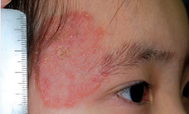
Facial dermatophytosis (tinea faciei) is a superficial infection of the smooth skin of the face, excluding the moustache and beard area, caused by dermatophytes. It is characterized by a well-defined erythematous patch with a scaly surface. ICD-10 Code: B35.0
In 60% of cases, the condition occurs in children under 12 years old. The age range described in the literature is from 8 days to 86 years. Peak incidence is between 2 and 14 years in children and between 20 and 40 years in adults. The disease is more common in women, with a ratio of 1.06:1 to men.
The exact prevalence is unknown. According to various studies, it ranges from 2 to 19% of all dermatophytoses.
The cause of the disease is infection with dermatophyte fungi. Infection can occur through:
- Direct contact with infected individuals or animals (cats, dogs).
- Contact with contaminated objects.
- Autoinoculation when the infection is localized on the trunk, limbs, or scalp.
The most commonly identified pathogens are anthropophilic and zoophilic fungi such as Trichophyton mentagrophytes, Trichophyton rubrum, Microsporum audouinii, Microsporum canis, Trichophyton tonsurans, Trichophyton verrucosum. Cases of Trichophyton arthroderma benhamiae infection in domestic guinea pigs have also been reported.
The disease is more common in children during the fall season when they return from vacations in rural areas where they come into contact with animals while playing. Facial dermatophytosis is a common infection among athletes, especially wrestlers (tinea gladiatorum), usually caused by T. tonsurans. In adults, risk factors include immunodeficiency and diabetes mellitus.Tinea faciei is characterized by the appearance of an erythematous patch with well-defined borders, most often covered with grayish-white scales. As it grows, the patch takes on a circular shape with resolution in the center and a raised red border at the periphery, on the surface of which papules, vesicles and pustules may be observed. The size varies from 1 to 10 or more centimeters in diameter.
The patch is most often solitary and asymmetrical. The most common sites are the cheeks, less commonly the periorbital area, forehead, nose, and chin. Patients often complain of itching and increased sensitivity to sunlight.The diagnosis is based on the medical history, clinical presentation, and laboratory data:
Microscopy of a specimen with KOH test reveals fungal hyphae.
Cultural examination allows identification of the causative organism and determination of its sensitivity to drugs.- Discoid lupus erythematosus
- Seborrheic dermatitis
- Rosacea
- Atopic dermatitis
- Contact dermatitis
- Candidiasis
- Polymorphous light eruption
- Acne vulgaris
- Perioral dermatitis
- Sarcoidosis
- Secondary syphilis
- Sweet syndrome
- Majocchi granuloma
- Drug-induced photoallergic reaction
- Granuloma Annulare. In Granuloma Annulare, papules and nodules merge, forming rings and semicircles. The elements often have a violet, not red, color, and there is no scaling.
- Nummular eczema presents as circular patches, but there are no clearing centers. The edge is not raised, and there are usually crusts on the patches without scaling.
- Psoriasis shows red papules or plaques, usually without clearing in the center. The scales are larger (they are smaller in body dermatophytosis). Scraping the scales leads to the appearance of pinpoint drops of blood (Auspitz's sign).
To prevent reinfection, the infection in domestic animals should be identified and treated.
Topical treatment is prescribed for 2-6 weeks.
- Terbinafine 1% cream - apply once or twice a day.
- Clotrimazole 1% cream - apply twice a day.
- Econazole cream - apply once or twice a day.
- Oxiconazole cream - apply twice a day.
- Ketoconazole cream - apply twice a day.
- Ciclopiroxolamine cream or lotion - apply twice a day.
- Terbinafine - 250 mg daily for 2 weeks.
- Itraconazole - 100-200 mg per day for 2-4 weeks or 200 mg twice a day for 1 week.
- Griseofulvin - 330-375 mg per day for 4-8 weeks.
- Fluconazole - 150 mg per week for 2-4 weeks.

