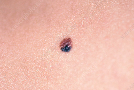
Dysplastic nevus is an acquired melanocytic lesion with atypical clinical and histological features. ICD-10 code: D23.9.
Dysplastic nevi are classified as familial (atypical nevus syndrome) and sporadic, depending on the presence of a family history of cutaneous melanoma. They are found in 5% of the Caucasian population in the United States and in 1.8-18% of the population in other countries. They are present in almost all patients with a family history of melanoma and in 30-50% of patients with sporadic cutaneous melanoma. They predominantly affect individuals of Caucasian race with no gender preference. Familial clustering has been documented. Most dysplastic nevi appear in childhood and adolescence, sometimes during periods of growth. Sporadic lesions may occur at any age, even up to the age of 60.
Familial nevi follow an autosomal dominant pattern of inheritance with relatively high penetrance. Several genes, including 1p36, 9p21, are considered as causal factors, suggesting a polygenic etiology. Sun exposure, especially intense and intermittent, is considered a contributing factor to the development of the condition. However, nevi can also appear on completely covered areas of the body.Dysplastic nevi typically appear later than acquired melanocytic nevi, just before the onset of puberty, and continue to appear for many years, even into old age. They are not characterized by involution (at least they undergo it much less frequently than acquired nevi). Unlike dysplastic nevi, acquired melanocytic nevi never appear in the elderly, and those that do tend to disappear with age.
Common characteristic features:
- Macular lesion with papular components.
- Asymmetry.
- Diameter exceeds that of commonly acquired nevi, typically ranging from 5 to 15 mm.
- Irregularly shaped and indistinct borders.
- Flat or bumpy surface with accentuated skin lines visible under lateral illumination.
- Uneven pigmentation with shades of dark brown, light brown, flesh-colored, pink, and blackish-brown.
- Erythema may be present within or around the lesion (halo).
- Round, oval, or ellipsoidal shape.
- Single or, more often, multiple (up to hundreds) randomly scattered lesions, especially in familial cases.
- Localization: trunk, hands, legs, dorsum of hands and feet, buttocks, and the hairy part of the head (both exposed and covered areas). Mucous membranes can also be affected.
Clinical subtypes:
- Fried egg type.
- Targetoid.
- Lentiginous.
- Seborrheic keratosis-like.
- Erythematous.
- Melanoma-like.
- Acquired melanocytic nevi
- Congenital melanocytic nevi
- Nevus spilus
- Spitz nevus
- Melanoma in situ/ melanoma
- Seborrheic keratosis
- Actinic keratosis
- Basal cell carcinoma
General therapeutic recommendations
Treatment depends on the number of nevi, the patient's history of melanoma, and family history (presence of atypical nevi and cutaneous melanoma in relatives).
Patients should undergo regular screening throughout their lives.
The evaluation should include:
- Examination of the entire skin surface.
- Creating a “mole-map” or diagram of the location of the nevi, which should be periodically updated.
Patients should be warned about the potential dangers of sun exposure and taught how to protect themselves and their children. They should be encouraged to avoid sunbathing and to use sunscreen. These patients should not tan or use tanning beds.
Close relatives should also be screened for the presence of dysplastic nevi and cutaneous melanoma and should be monitored regularly.
Clinically typical nevi do not require routine excision. Patients with a few nevi should be monitored annually or every six months if there is a family history of cutaneous melanoma.
Lesions that require excision include:
- All lesions suspicious for skin melanoma.
- Changing lesions, for example, those increasing in size or changing in pigmentation, shape, and/or border.
- Lesions that are difficult for the patient to monitor independently (e.g., on the scalp, genitalia, upper back), or if there are doubts that the patient will attend for follow-up examinations.
- Lesions in the areas, such as palms, soles, and nail beds.
Complete surgical excision is the treatment of choice. Excision should always be followed by histologic examination to rule out malignancy.
Avoid laser, electrosurgery, cryotherapy, and other treatment options that result in physical destruction of the lesion, as these methods do not allow histologic examination of the lesion.
