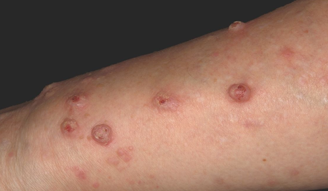
Keratoacanthoma is a rapidly growing epidermal tumor that originates from hair follicles and occurs predominantly on exposed areas of the body and extremities, especially the extensor surfaces. ICD-10 Code: L85.8.
Etiology and epidemiology
The disease occurs between the ages of 14 and 90, with a peak incidence between the ages of 45 and 70, and 80% of patients are over the age of 40. An exception is Ferguson Smith multiple keratoacanthoma, which occurs in children and adolescents. Males are three times more likely to be affected than females.
Etiology and pathogenesis are unknown. The viral nature of keratoacanthoma remains controversial, although human papillomavirus DNA has been detected in tumor tissue in nearly half of cases. Multiple self-resolving keratoacanthomas of Ferguson-Smith are inherited in an autosomal dominant pattern. Genetic predisposition may also be a factor in other forms of the tumor.
Predisposing factors include:
- Ultraviolet radiation
- Immune disorders: In individuals treated with cyclosporine, infliximab, those who have undergone radiation therapy, or those who have had accidental radiation exposure.
- Trauma: Development of tumors around surgical wounds, burn scars, postoperative scars, tattoos (especially red ink).
- Smoking: Particularly relevant when localized in the oral mucosa.
- Contact with chemical substances: Insecticides, petroleum distillation products, tar.
Keratoacanthoma is believed to arise from hyperplastic epithelium within one or more closely situated hair follicles and associated sebaceous glands. The occurrence of keratoacanthoma on the mucous membranes of the oral cavity is thought to be related to ectopic sebaceous glands.
According to the WHO International Classification of Skin Tumors (2018), keratoacanthoma is classified as a type of squamous cell carcinoma.Solitary Keratoacanthoma
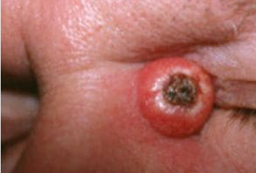
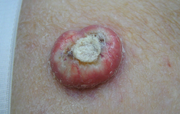
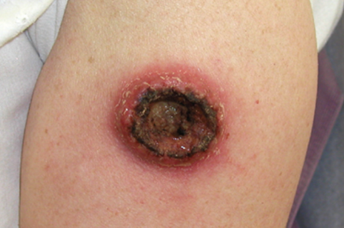
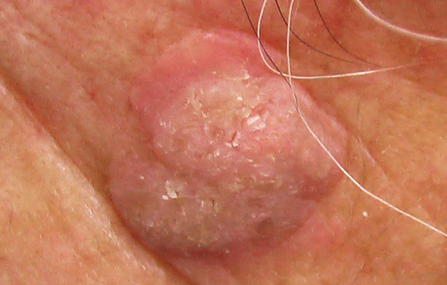
The most common type begins as a small, solitary, firm, hemispherical papule of skin color that rapidly grows into a round or oval exophytic nodule on a broad base. The color is reddish, sometimes with a bluish tinge, or normal skin color, with a size of 0.5-3 cm in diameter. A molluscum-like appearance is characteristic. A crater-like depression filled with loose or dense brownish keratinous masses (pseudoulcers) forms in the center of the tumor. The peripheral high rim surrounding the keratin plug has a smooth surface and appears pink or skin-colored. Keratotic masses are easily removed to reveal underlying papillomatous growths. Typically a keratoacanthoma is a solitary lesion, but in some cases multiple (up to 10) lesions may be present.
The usual localization is on sun-exposed areas (head, hands, forearms, and less commonly, upper trunk and lower extremities). Rare sites include the genitals, oral mucosa (cheeks, hard palate, tongue, gums), conjunctiva, and periocular area. On the conjunctiva, keratoacanthoma appears as a white nodule with central hyperkeratosis and surrounding dilated episcleral vessels.
The active growth phase may be followed by a stabilization phase during which the size of the tumor does not change. Typically, spontaneous regression occurs within 3-9 months with disappearance of the tumor nodule and formation of an atrophic scar. In some cases, the stabilization phase does not occur and the tumor can reach giant sizes (up to 10-20 cm in diameter).
It has been reported that squamous cell carcinoma occurs in 5-6% of cases with metastasis to parathyroid glands and regional lymph nodes.Giant Keratoacanthoma
Keratoacanthoma centrifugum marginatum
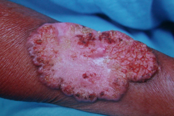
A rare variant characterized by progressive horizontal growth with central healing. It begins as small papules that slowly evolve into annular or polycyclic patches with raised margins, sometimes with papulo-nodular elements. Dense pigmented mini-papules may be present in the center of the scarred patches. Such keratoacanthomas are predominantly solitary and can sometimes reach 20 cm or more in diameter. In some cases, the tumors are multiple, unilateral or bilateral, primarily on the extremities, often the lower extremities, although localization can be anywhere. Spontaneous regression occurs within 6-12 months, but in some cases it may last for several years. Cases have been described in which keratoacanthoma centrifugum marginatum coexisted with giant and multiple tumors.
The tumor can simulate annular elastolytic giant cell granuloma, elastosis perforans serpiginosa, discoid lupus erythematosus, squamous cell carcinoma, tuberculosis, syphilis, deep mycoses.Keratoacanthoma en plaque
Subungual Keratoacanthoma
Persistent Keratoacanthoma
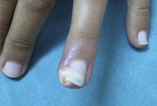
A rare, aggressive form seen primarily in middle-aged men. It is often preceded by trauma. The first finger of the hand is most commonly affected. The tumor is characterized by rapid growth, pain, and destruction of the underlying bone. The nodule becomes visible after detachment of the nail plate. It is similar to a wart and squamous cell carcinoma. Spontaneous involution is rarely seen.
Recurrent Keratoacanthoma
A rare, aggressive form seen primarily in middle-aged men. It is often preceded by trauma. The first finger of the hand is most commonly affected. The tumor is characterized by rapid growth, pain, and destruction of the underlying bone. The nodule becomes visible after detachment of the nail plate. It is similar to a wart and squamous cell carcinoma. Spontaneous involution is rarely seen.
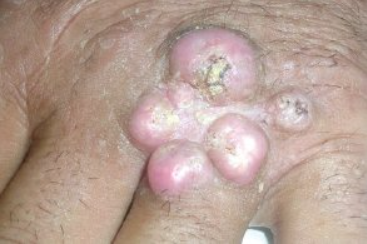
Multinodular Keratoacanthoma
Multiple keratoacanthoma Ferguson Smith type
Generalized Eruptive Keratoacanthomas of Grzybowski
Multiple Familial Keratoacanthoma of Witten and Zak
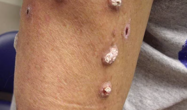
An intermediate type between Ferguson-Smith and Grzybowski keratoacanthomas. The hereditary nature of the disease is presumed. It is characterized by simultaneous eruption of multiple milia-sized and larger self-resolving elements. In some cases, nodular-ulcerative formations of medium and large size and eruptions on the mucous membrane of the oral cavity are observed.
Keratoacanthomas in Other Diseases
Solitary and multiple keratoacanthomas can also be observed in Muir-Torre syndrome and xeroderma pigmentosum.
Diagnosis is based on clinical manifestations; excisional biopsy is performed for solitary or multiple lesions; diagnostic biopsy of the raised margin is performed for large lesions.
Histopatology: Considered by some to be a highly differentiated form of squamous cell carcinoma, histological examination reveals a circumscribed proliferation of well-differentiated keratinocytes. This has been described as multilobular exophytic or endophytic cyst-like invagination of the epidermis. The epidermis extends over the tumor, and there is a central horn plug of keratin. Peripheral to the keratin-filled crater are lip-like, peripheral borders of the epidermis. Intraepidermal neutrophilic abscesses are visualized in addition to horn pearls. The cells of the keratoacanthoma tumor are enlarged and atypical keratinocytes. They have a cytoplasm described as eosinophilic. Due to 3 stages of solitary keratoacanthoma, the histological examination can vary between stages. In comparison to squamous cell carcinoma, keratoacanthoma has intraepidermal microabscesses and tissue eosinophilia more commonly found. Recently keratoacanthoma has been reclassified as squamous cell carcinoma keratoacanthoma type (SCC-KA).
Cite: Zito PM, Scharf R. Keratoacanthoma. [Updated 2023 Mar 7]. In: StatPearls [Internet]. Treasure Island (FL): StatPearls Publishing; 2023 Jan-. Available from: https://www.ncbi.nlm.nih.gov/books/NBK499931/
- Squamous cell carcinoma
- Common warts
- Giant molluscum contagiosum
- Actinic keratosis
- Cutaneous horn
- Basal cell carcinoma.
- The multiple form should be differentiated from Darier's disease.
Due to the spontaneous involution of keratoacanthomas, waiting for 3-6 months is possible.
For solitary tumors, surgical excision, curettage, electro-, cryo-, or laser destruction (CO2, Er:YAG, Argon laser) are performed.
Alternative methods include:
- 5-fluorouracil cream
- Pirospidine cream
- Imiquimod
- Intralesional injection of methotrexate, interferon-alpha 2a
Due to the aggressive nature of subungual keratoacanthomas, amputation of the distal phalanx may sometimes be necessary.
For recurrent cases, surgical excision using the Mohs technique, oral administration of acitretin, isotretinoin, low doses of methotrexate for a short course is recommended.In keratoacanthoma centrifugum marginatum , after surgical excision, reconstructive procedures may be necessary, retinoids are used for up to 5-6 months, and sometimes radiotherapy is prescribed.
Good results are observed in multiple keratoacanthomas of Ferguson-Smith type with intravenous administration of 5-fluorouracil (12 mg/kg/day in 5-day cycles with 2-day intervals for 6 weeks).
For Grzybowski and Witten-Zak keratoacanthomas, the use of retinoids is preferred: Acitretin oral 50-75 mg once daily for 4-11 weeks.
If there is no effect, cyclophosphamide is prescribed (200 mg/day for 3 months, followed by dose reduction to 100 mg, continued for another month; good effect was observed with pulse therapy - 1 g/month for 6 months).
In recent years, photodynamic therapy and EGFR tyrosine kinase inhibitor - erlotinib are being tested for various variants of keratoacanthoma.
Prognosis: Keratoacanthoma is a rapidly growing neoplasm that often regresses spontaneously (usually within 2-6 months). In some cases, can be transformation to squamous cell carcinoma.
