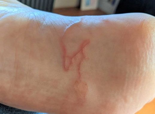
Cutaneous larva migrans is a parasitic disease caused by the larvae of intestinal nematodes, most commonly hookworms, characterized by the appearance of pruritic, migrating serpentine lines on the skin. ICD-10 Code: B76.9
The most common causes of the disease are hookworms (Ancylostoma braziliense, Ancylostoma ceylonicum, Ancylostoma caninum, Ancylostoma duodenale) - intestinal parasites of animals, especially dogs, and occasionally humans. Eggs of these helminths, found in the feces of infected hosts, mature in a moist and warm environment, such as sandy soil in shaded areas, releasing small, transparent larvae up to 1 cm in length.
Upon contact with contaminated environments, the larvae rapidly penetrate intact skin and cause a local inflammatory response due to the products they secrete as they migrate. The disease is most commonly contracted by people whose occupations involve contact with warm, moist, sandy soil, such as farmers, gardeners, plumbers, electricians, carpenters, fishermen, hunters, as well as beachgoers and children playing in the dirt. For most helminths, humans are a "dead-end" host: larva die without reaching sexual maturity, and the disease resolves on its own.Pruritus at the site of larval penetration occurs within hours of infection, with typical lesions appearing 2-3 weeks later. Patients notice pruritic, inflamed, serpiginous patches that migrate in a snake-like manner. Once under the skin, larva move laterally through the epidermis, covering a distance of a few millimeters to several centimeters per day, creating sinuous, slightly elevated intradermal tracks that are 2-3 mm wide and 4 to 30 cm long. These tracks, filled with serous fluid, appear as urticarial stripes, varying in color from pinkish red to purplish violet, resembling the track of a sea snail moving aimlessly on the sand at low tide. Larva is usually located 0.5-3 cm in front of the visible end of the track in the reactive zone.
Several larvae may be present in a single area, forming several closely spaced wavy lines. The most commonly affected areas are the soles and ankles, followed by the buttocks, genitals, and palms. Itching ranges from mild to severe, and secondary infection and eczematous inflammation may sometimes occur.
If left untreated, larva die within 2-8 weeks, although rare cases of persistence for up to 1 year have been reported. In rare cases, the larvae may emerge spontaneously as the epidermis matures.
Clinical Forms:
Larva currens (cutaneous strongyloidiasis): Caused by Strongyloides stercoralis, larva exhibit rapid movement (approximately 10 cm/h). Lesions at the site of larval penetration include papules, papulovesicles, and urticaria accompanied by intense pruritus. They are commonly found on the perianal area, buttocks, thighs, back, shoulders, and abdomen. Larvae migrate from the skin into the bloodstream, causing the itching and skin manifestations to disappear. The worm reproduces in the intestinal mucosa.
Larva migrans syndrome (visceral form): Migrating larvae of canine and feline roundworms (Toxocara canis, Toxocara cati) and human ascarids (Ascaris lumbricoides) infest internal organs. Symptoms include persistent eosinophilia, hepatomegaly, and occasionally pneumonia.
Loeffler's syndrome may be a complication of Ancylostoma braziliense infection, resulting in localized pulmonary infiltrates and eosinophilia.- Scabies
- Schistosomiasis
- Swimmer's itch(cercarial dermatitis)
- Portuguese man o' war sting
- Jellyfish contact
- Erythema annulare centrifugum
- Erythema migrans (Lyme disease)
- Phytophotodermatitis
- Contact dermatitis
- Tinea pedis (Athlete's foot)
- Myiasis
- Loiasis
- Dracunculiasis
- Gnathostomiasis
- Foreign body
In typical cases, migration and itching reduce within 2-3 days after the start of treatment.
Systemic Therapy:
- Ivermectin: 200 mcg/kg (average dose 12 mg) as a single oral dose. Lesions heal within 5 days after starting ivermectin. A second course in the same dose is administered in case of relapse. Some authors recommend ivermectin for 10-12 days.
- Albendazole: Either 400 mg once daily orally or 200 mg twice daily orally for 3-7 days. It acts quickly, with itching disappearing within 3-5 days, and skin lesions resolving after 6-7 days of treatment.
- Thiabendazole: Administered orally at a dose of 50 mg/kg/day every 12 hours for 2-5 days. Maximum daily dose: 3 g. In the case of secondary infection, antibiotics are used. Topical or systemic steroids may be needed for severe itching.
Local Therapy:
Freezing the moving end of the lesion with liquid nitrogen is often ineffective.
Apply 10-15% thiabendazole as a liquid or cream topically 3-4 times daily for 5 days. It should be applied to the affected areas and 2 cm beyond the leading edge of the clinical lesion, as the parasite often resides beyond its borders.
