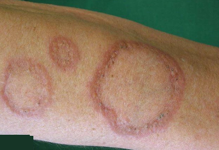
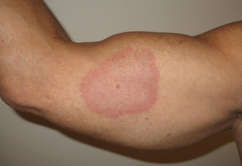
Granuloma Annulare is a benign skin disorder characterized clinically by annular papules and pathomorphologically by granulomatous inflammation. ICD-10 Code: L92.0.
The prevalence of granuloma annulare is estimated at 0.1-0.4% of the total number of patients with dermatological pathology. The etiology and pathogenesis of the disease are unknown. The disease is considered to be multifactorial, with a certain role attributed to chronic infections (tuberculosis, rheumatism, chronic respiratory infections), sarcoidosis, endocrine disorders, diabetes (more common in the generalized form of the disease), prolonged use of medications (vitamin D). Sometimes there is an association with autoimmune thyroiditis. Trauma may also play a provoking role in the development of granuloma annulare. An association between granuloma annulare and tuberculin skin test and Bacillus Calmette-Guérin (BCG) vaccination has been described. Viral infections (HIV infection, Epstein-Barr virus, herpes simplex virus, and varicella-zoster virus) may also contribute to the development of the disease. Granuloma annulare associated with immunodeficiency (HIV infection, post-liver transplantation) is more common.
Currently, the following mechanisms are considered in the development of the dermatosis:
- Microangiopathies leading to degradation of connective tissue.
- Primary degenerative process in connective tissue with a granulomatous reaction.
- Lymphocyte-mediated immune reaction activating macrophages.
Based on the clinical presentation of the disease, the following forms are distinguished:
- Localized granuloma annulare.
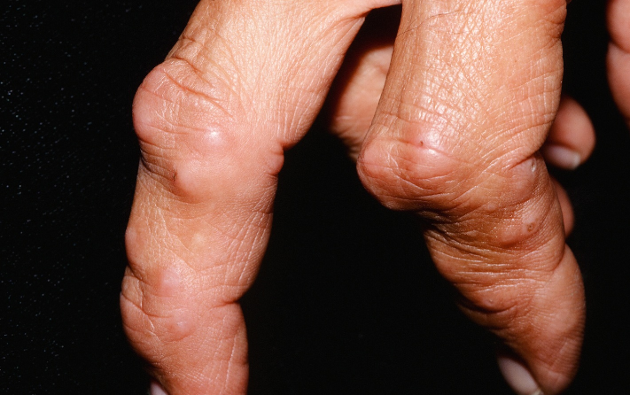
- Deep (subcutaneous) granuloma annulare.
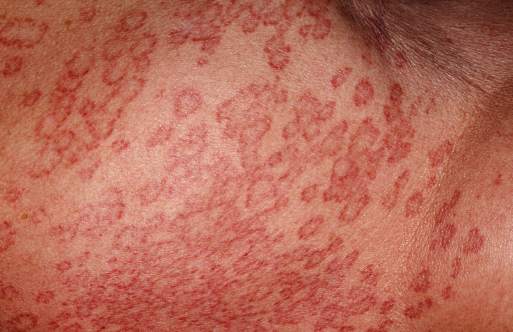
- Disseminated (generalized) granuloma annulare.
- Perforating granuloma annulare.
- Patch granuloma annulare.
Localized Granuloma Annulare
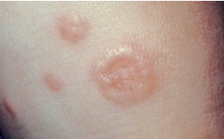
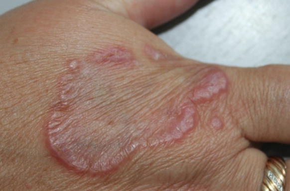
Localized Granuloma Annulare is the most common form of the disease and is primarily observed in children (90% of cases) and young individuals. It is characterized by the appearance of small (0.1-0.5 cm in diameter), firm, smooth, hemispherical papules of pink, red, livid, or normal skin color. These papules are typically arranged in a circle or in groups forming a semi-annular pattern and are commonly found on the backs of the hands, feet, shins and forearms (60% on hands and arms, 20% on feet and shins). Less commonly, it affects the neck, trunk, wrists, ankles, periorbital area, eyelids, scalp, and occasionally the palms and soles. Cases of genital localization have been reported.
The peripheral zone of annular eruptions consists of multiple discrete or confluent papules, macules, or nodules with a smooth surface that is reddish, livid, or flesh-colored. The peripheral zone has a moderately firm consistency to palpation. The skin within the annular eruptions may have a purplish or pigmented appearance. Isolated firm papules and nodules may be present in addition to the annular elements.
In the papular variant, the configuration of the eruptions is not circular and the papules often have a central dimple, especially on the hands, and are isolated from each other.
There are no subjective sensations. The lesions gradually increase in diameter from 1 to 5 cm or more. Over time, they may partially resolve or recur at the same site. The localized form may be associated with the subcutaneous and patch-like variants of granuloma annulare.Deep Granuloma Annulare
Disseminated Granuloma Annulare
Perforating Granuloma Annulare
Patch granuloma annulare
The diagnosis of granuloma annulare is primarily based on clinical presentation; however, in certain cases (suspected disseminated and deep forms of the disease), histopathologic examination of skin biopsies is necessary.
Referrals to other specialists are indicated depending on the circumstances: consultation with an general practitioner (if physiotherapy is prescribed, it is mandatory), endocrinologist, infectious disease specialist, otolaryngologist and pulmonologist may be necessary.- Erythema elevatum diutinum
- Tuberculoid type of leprosy
- Papular form of sarcoidosis
- Necrobiosis lipoidica
- Rheumatic nodules
- Lichen planus
- Psoriasis
- Tinea corporis
Treatment Objectives
- Resolution of eruptions
- Prevention of recurrences
General Notes:
- When planning therapy, it's important to consider the tendency of granuloma annulare to spontaneously resolve. In approximately 75% of cases, lesions spontaneously resolve within 2 years. Although the recurrence rate reaches 40%, new lesions may also disappear spontaneously. If necessary, correction of carbohydrate metabolism is performed, along with treatment of any concomitant pathology (tuberculosis, diabetes).
- For localized granuloma annulare, topical glucocorticosteroids are the preferred treatment. For disseminated skin involvement, systemic medications or phototherapy are used in combination with topical therapy.
Topical Therapy
Glucocorticosteroids:
- Hydrocortisone 17-butyrate, cream, ointment 0.1%, once daily in the evening for 14 days, then once every 2 days for 2-3 weeks.
- Alclometasone dipropionate, cream, ointment 0.05%, once daily in the evening for 14 days, then once every 2 days for 2-3 weeks.
- Betamethasone dipropionate, cream, ointment 0.025%, 0.05%, once daily in the evening for 14 days, then once every 2 days for 2-3 weeks.
- Betamethasone valerate, cream, ointment 0.1%, once daily in the evening for 14 days, then once every 2 days for 2-3 weeks.
- Methylprednisolone aceponate, cream, ointment, emulsion 0.1%, once daily in the evening for 14 days, then once every 2 days for 2-3 weeks.
- Mometasone furoate, cream, ointment, lotion 0.1%, once daily in the evening for 14 days, then once every 2 days for 2-3 weeks.
- Clobetasol propionate, cream, ointment 0.05%, once daily in the evening for 14 days, then once every 2 days for 2-3 weeks.
Systemic Therapy
- Tocopheryl acetate: for children aged 3-10 years, 50-100 mg orally per day; for children older than 10 years, 100-200 mg orally per day; for adults, 200-400 mg orally per day for 20-40 days. (*Need more investigations)
- Vitamin E + retinol: 1 tab 1-3 times daily orally for 1 month. (*Need more investigations)
- Vitamin C + rutin: for children under 5 years, 1-2 tablets daily orally; for children aged 5-10 years, 1 tablet twice daily orally; for children older than 10 years and adults, 1 tablet 3 times daily orally for 20-40 days. (*Need more investigations)
Destructive Therapy
- Cryotherapy: once every 7-10 days, 3-5 procedures per lesion. The entire surface of small lesions and the active edges of larger lesions (diameter over 4 cm) are treated. Temporary side effects (pain, blister formation, local edema) and long-term complications (localized hypopigmentation and peripheral hyperpigmentation) are possible.
Treatment for Pregnant Patients:
There is no data on treating granuloma annulare in pregnant individuals. In such clinical situations, the use of local therapy methods is permissible:- Topical application of tocopheryl acetate (vitamin E) twice daily, with one application under occlusion, for 2 weeks. (Need more investigations)
- Cryotherapy: once every 7-10 days, 3-5 procedures per lesion.

