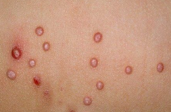
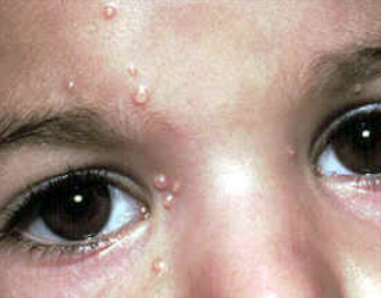
Molluscum Contagiosum is a benign viral skin condition characterized by the appearance of dome-shaped nodules on the skin, occasionally on mucous membranes, ranging in size from a pinhead to a small pea, with a central umbilicated depression. ICD-10 Code: B08.1.
The disease is caused by Orthopoxvirus, which belongs to the family Poxviridae, subfamily Chordopoxviridae, and genus Molluscipoxvirus. There are four types of Molluscum contagiosum virus: MCV-1, MCV-2, MCV-3, and MCV-4. The most common type is MCV-1; MCV-2 is typically found in adults and is transmitted through sexual contact. Orthopoxvirus is a DNA-containing virus that cannot be cultured in chicken embryo tissue and is not pathogenic in laboratory animals. The disease is widespread and affects people of all ages.
Infection occurs through direct contact with an infected person or carrier, or indirectly through personal and household items. The incubation period of the disease varies from 1 week to several months, with an average of 2 to 7 weeks. The disease is most commonly diagnosed in children between the ages of 1 and 4. In older children, infection typically occurs during swimming pool visits or contact sports. Children with eczema or atopic dermatitis treated with glucocorticosteroids are more susceptible.
In young adults, molluscum contagiosum is often sexually transmitted. In middle-aged and elderly individuals, prolonged use of glucocorticosteroids and cytostatics may be a predisposing factor for the disease.
In HIV-infected patients, due to the immunodeficient state of the body, there is an increased susceptibility to molluscum contagiosum, which is characterized by a recurrent course. The prevalence of the disease ranges from 1.2% to 22% of the population in different countries.Molluscum Contagiosum lesions can be found on any part of the skin. In children, the lesions are more commonly found on the skin of the face (often on the eyelids and forehead), neck, upper chest (especially in the armpits), upper extremities (backs of the hands); in adults, they are found on the lower abdomen, pubic area, inner surface of the thighs, skin of the external genitalia, and around the anus. Eyelid involvement may be associated with conjunctivitis. In HIV-infected individuals, lesions are more frequently localized on the face, neck, and trunk.
The lesions of molluscum contagiosum are small nodules, 0.1-0.2 cm in size, with a hemispherical or slightly flattened shape. They are firm, painless, the color of normal skin or pale pink, often with a waxy sheen and a central umbilication. Papules rapidly increase in size to 0.5-0.7 cm, are isolated on unaffected skin, and are occasionally surrounded by a mildly inflammatory rim. When compressed laterally, papules release a white, cottage-cheese-like material from the central opening, consisting of degenerated epithelial cells with large protoplasmic inclusions. The number of lesions may vary from 5-10 to several dozen or more.
In majority of cases, the eruptions are not accompanied by subjective sensations and represent only a cosmetic problem for the patient. Typically, the condition is self-limiting and the morphologic elements may disappear spontaneously without treatment. However, in children, molluscum contagiosum may have a prolonged course (from 6 months to 5 years) as a result of autoinoculation of the pathogen.
Phenomenas in Molluscum Contagiosum:
- Koebner Phenomenon. Linear arrangement of rash elements due to autoinoculation of the molluscum virus.
- Halo Phenomenon. Appearance of a white halo around papules, likely associated with the secretion of a protein that inhibits inflammation around the lesion.
- Meyerson's Phenomenon (Molluscum Dermatitis). Development of eczematous reactions, probably representing an immune-mediated response of the body to the molluscum virus and a precursor to regression.
- Flat Warts
- Common Warts
- Keratoacanthoma
- Milia
- Closed Comedones
- Cryptococcosis
- Herpes simplex virus
- Bacterial folliculitis
- Sebaceous hyperplasia
Treatment Goals
- Regression of the rash.
- Prevention of recurrences.
General Notes on Therapy
The primary goal of therapy is to destroy molluscum contagiosum lesions. Because of the potential for autoinoculation, it is essential that all lesions be removed. Prior to initiating therapy, a thorough examination of the patient's entire skin surface should be performed, paying particular attention to skin folds. Patients should be advised not to shave areas with lesions as this may lead to autoinoculation.
Topical treatment
In 1999, Brazilian dermatologist Ricardo Romiti introduced a painless and effective method of treating molluscum contagiosum using a 5% aqueous solution of potassium hydroxide. Its safety and efficacy, up to 92%, were subsequently confirmed by numerous clinical trials in accordance with the principles of evidence-based medicine. Various concentrations of the treatment have been proposed, ranging from 2.5% to 20%, but the optimal concentration has been found to be 5%.
The essence of the method is to apply the solution to the affected areas twice a day using a special pipette, brush or simply a cotton swab until a reddish halo forms around the lesion. In some cases, a small erosion may develop at the site of the molluscum, which heals quickly without scarring. The redness typically begins within 7-14 days of the start of the procedure. Treatment should be discontinued at this time. Complete disappearance of the rash is observed within 4 weeks.
Products containing a 5% potassium hydroxide solution include the British product "MolluDab" and the French product "Molutrex".
Destruction Methods
- Curettage
- Cryotherapy
- Laser therapy of molluscum contagiosum lesions: Using a CO2 laser or a pulsed dye laser
Local anesthesia is used to reduce pain and discomfort during destruction of molluscum contagiosum lesions.
After destruction of molluscum contagiosum lesions, the skin areas where they were located are treated with antiseptics
Special situations
Patients with atopic dermatitis have a high risk of scarring with a large number of lesions; therefore, curettage is undesirable. It is recommended to treat exacerbations of atopic dermatitis prior to initiating therapy for molluscum contagiosum.
In cases where molluscum contagiosum lesions are found in immunocompromised patients, treatment methods that disrupt skin integrity should be avoided, as these patients are at high risk of developing infectious complications. Cases of regression of molluscum contagiosum lesions after initiation of antiretroviral therapy have been reported.
All methods of destruction are permitted during pregnancy.
Treatment Outcome Requirements
- Resolution of lesions.
- Complete clinical remission.
Preventive Measures
- Preventive measures include isolation of affected children from groups until complete recovery and adherence to personal and public hygiene rules. During the treatment period, visits to swimming pools, sports facilities and public baths are prohibited.
- Preventive measures for molluscum contagiosum also include preventive screening of children in preschool institutions and schools, early detection of cases of molluscum contagiosum, timely treatment of affected individuals and their sexual partners.
- A person with molluscum contagiosum should use only their personal belongings and utensils until the end of treatment, avoid sexual and close physical contact, and refrain from visiting swimming pools or saunas.


