Lentigo

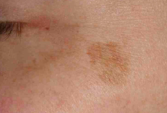
Solar lentigo 1

Lentigo - solitary or widespread benign pigmented spots that develop on sun-exposed areas of the skin. ICD-10 code: L81.4

There are three types of localized hyperpigmentation: freckles (ephelides), juvenile lentigo, and solar lentigo. All three types of spots are similar in size, distribution, and clinical appearance. They differ in the age of onset, clinical course, and response to light exposure.

Freckles appear in childhood and are inherited in an autosomal dominant manner. They are typically localized on the skin of the face, hands, and upper trunk. Their number increases and their color darkens in response to sun exposure. Without sun exposure, freckles become lighter. During winter, they often completely disappear. Juvenile lentigo is very common among individuals of white ethnicity.

Juvenile lentigo appears in childhood, and the average number in a prepubescent child is around 30. These spots do not increase in number or size and do not darken upon sun exposure. Juvenile lentigo can also be a characteristic feature of certain inherited syndromes.

Solar lentigo is most often found on sun-exposed areas of the skin in individuals of Caucasian ethnicity. With age, their number and size increase. Approximately 75% of Caucasians over the age of 60 have one or more spots. These spots develop in response to actinic damage.

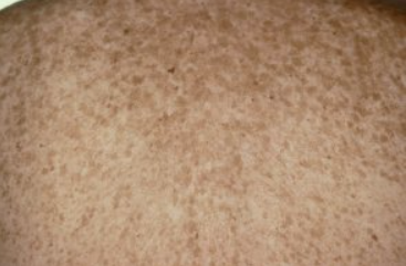
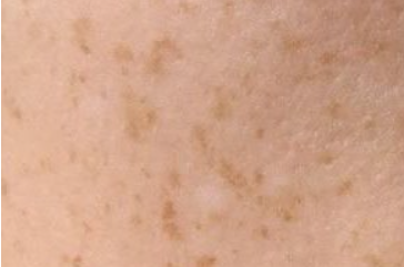
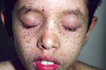
Freckles (ephelides)


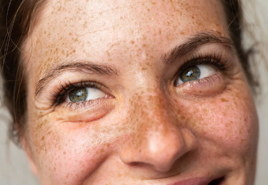
Freckles
They are well defined, 1-2 mm in diameter and uniform in color. The color of freckles varies from red or tan to light brown. The number of freckles can range from a few rare spots on the nose and cheeks to hundreds. They are typically localized to the skin of the face, hands, and upper trunk.

Juvenile Lentigo (Lentigo Simplex)


Lentigo simplex
They are round or oval spots 2-10 mm in diameter. They are usually darker than freckles and have a uniform color that resembles tan, brown, or black. Their color is consistent, although the pigmentation may have a lacy or granular pattern. A drop of mineral oil applied to the spot reduces surface shine and helps to assess the type of pigmentation.

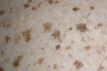
Solar Lentigo


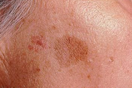
Solar lentigo 2
Large oval or geometric spots ranging in size from 2 to 20 mm. The color is often uniform, although the pigmentation may appear granular. Multiple lentigines resemble ink spots, although their borders are clearly defined. Signs of actinic damage are visible on the surrounding skin. Solar lentigo appears on sun-exposed areas of the face, hands, dorsal side of the hands, and upper trunk after sun exposure or many years of sun exposure. The number and size of these flat brown spots increase over time.

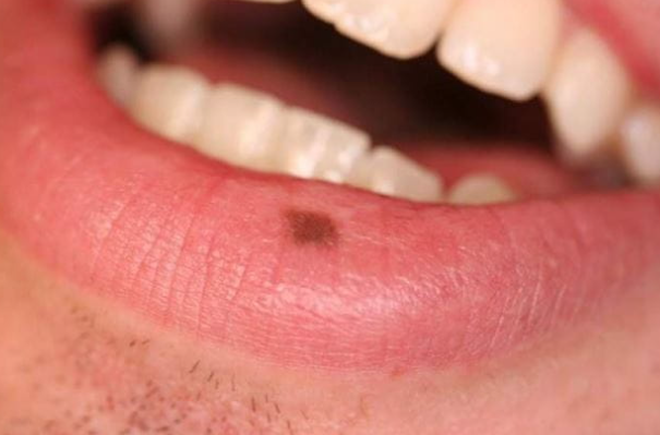
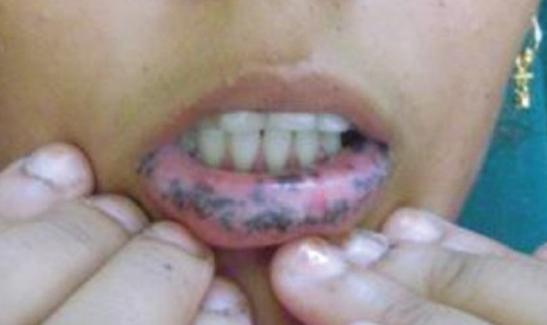
Labial Melanotic Macule


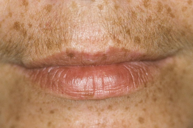
Labial Melanotic Macule
These brown spots on the red border of the lower lip have a smooth border and are benign.

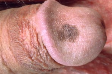
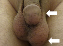
Penile Lentiginosis


Penile Lentiginosis
Often one, occasionally several brown spots on the shaft, glans, and foreskin of the penis.

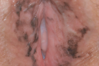
Vulvar Lentigo (Vulvar Melanosis)


	Vulvar Melanosis
Often one, occasionally several brown, dark brown, and/or black spots on the labia majora and labia minora.

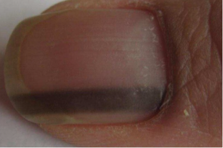
Nail Lentigo


Nail Lentigo
Nail appears with stripes (longitudinal melanonychia).

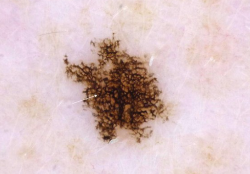
Reticulate Black Solar Lentigo (Ink-Spot Lentigo)


Ink-Spot Lentigo
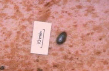
Appears as a dark brown or black, often solitary spot with noticeably irregular borders. It has a reticulated structure and resembles an "ink spot" on the skin.

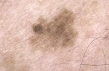
Atypical Solar Lentigo


Atypical Solar Lentigo
Appears as a large solitary spot with irregular shape, with multicolored (ranging from tan to black) pigmentation and irregular structure. Unlike regular solar lentigo, atypical lentigo has areas of melanocyte spread.

Multiple Lentigines (Lentiginosis)


Multiple Lentigo
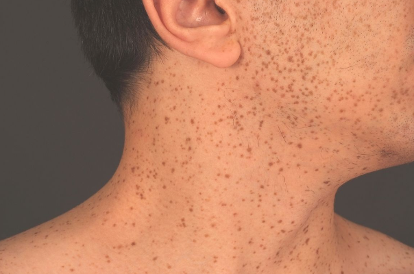
Characterized by multiple eruptions not associated with systemic disorders, but may be a sign of an autosomal dominant syndrome.

PUVA-Induced Lentigines


PUVA-Induced Lentigines
Develop in 1.5-4% of patients undergoing UVA therapy. They are typically generalized dark brown spots, including palms and soles, resulting from melanocyte hypertrophy and hyperplasia.

Peutz-Jeghers Syndrome


Peutz-Jeghers Syndrome
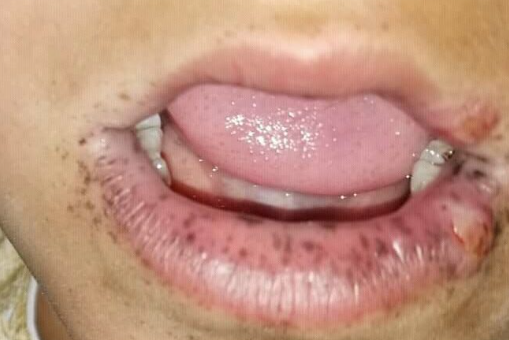
It is associated with intestinal polyposis and involves multiple lentigines on the oral mucosa.

LEOPARD Syndrome (Noonan syndrome with multiple lentigines)


LEOPARD Syndrome
Includes: Lentigines - multiple lentigines; Electrocardiogram abnormalities - EKG deviations; Ocular disorders - eye pathology; Pulmonary stenosis - narrowing of the pulmonary artery; Abnormalities of genitalia - genital pathology; Retardation of growth - growth delay; Deafness - hearing impairment.

Carney Complex


Carney Complex
Known by the acronym LAMB or NAME. This syndrome consists of lentigines, atrial and/or mucocutaneous myxomas, myxoid neurofibromas, freckles, and blue nevi.

Cronkhite-Canada Syndrome


Cronkhite-Canada Syndrome
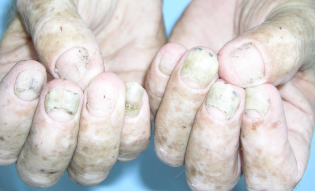
Lentigines on the mucous membrane of the cheeks, face, palms, and soles, along with alopecia, onychodystrophy, and intestinal polyps.

Laugier-Hunziker Syndrome


Laugier-Hunziker Syndrome
Characterized by dark brown spots up to 4-5 mm in diameter, mostly on the mucous membrane, sometimes extending to the red border of the lips. In some patients, these elements are found on the genitals. Nail changes in form of melanonychia are observed in 2/3 of cases.

Cantu Syndrome


Cantu Syndrome
Cantu Syndrome (Hyperkeratosis-Hyperpigmentation Syndrome) characterized by small areas of hyperpigmentation primarily resulting from sun exposure, as well as pinpoint papular hyperkeratosis on the palms and soles.

Bannayan-Riley-Ruvalcaba Syndrome


Bannayan-Riley-Ruvalcaba syndrome
Characterized by multiple malformations: macrocephaly, multiple lipomas, intestinal polyps, vascular malformations, and abnormal skin pigmentation on the penis.

Centrofacial Lentiginosis


Touraine Syndrome
Centrofacial Lentiginosis (Touraine Syndrome) - lentigines in the central part of the face: forehead, nose, occasionally on the lips and neck. Mucous membranes are not affected. It is associated with dysraphic status (gothic palate, cranial deformity, lack of incisors, sunken chest, spina bifida), neuro-psychiatric disorders (intellectual disability, epileptiform seizures, enuresis), and congenital hypertrichosis.

Diagnosis is usually based on clinical presentation, history, and dermoscopy.

Any lentigo with highly irregular borders or localized pigmentary increase or thickening should be biopsied to rule out melanoma and malignant lentigo.

  • Seborrheic keratosis
  • Pigmented actinic keratosis
  • Lentigo Maligna
  • Melanoma
  • In case of multiple lentigines, an associated syndrome can be suspected, such as Peutz-Jeghers syndrome, LEOPARD syndrome, or NAME syndrome (LAMB).
  • CafĂ©-au-lait spots
  • Melanocytic nevus
  • Melasma

  • Freckles do not require treatment and fade during the winter months.
  • Sunscreens prevent the appearance of new freckles and their seasonal darkening.
  • Juvenile lentigo also does not require treatment.
  • Prevention of solar lentigo is best achieved through sun protection measures, such as avoiding sun exposure, wearing hats, protective clothing, and using sunscreen.
  • Hydroquinone solutions, tretinoin, azelaic acid creams, and glycolic acid peels reduce hyperpigmentation over weeks and months.
  • Superficial cryosurgery shows the effectiveness