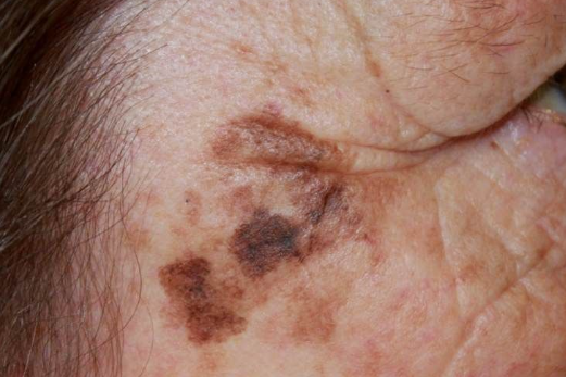
Lentigo maligna

lentigo maligna 1

Lentigo maligna is characterized by a distinctive, single, nonpalpable lesion with uneven brown-black pigmentation. It occurs on sun-exposed areas of the body, primarily the face, in elderly individuals and is considered a specific type of in situ melanoma arising from epidermal melanocytes. ICD-10 Code: D03.9

Published data indicate that lentigo maligna occurs in individuals over 40 years old, with a median age of onset 65 years. Blacks and Asians, including those from Southeast Asia, are rarely affected. The highest incidence is in fair-skinned individuals with skin types I, II, and III. The incidence increases with age, peaking in the seventh and eighth decades of life. Descriptive epidemiology suggests that the disease results from chronic cumulative sun exposure. Lesions typically appear on sun-exposed areas, primarily in individuals with fair skin.

Lentigo maligna typically presents as a single lesion, a hyperpigmented spot, on sun-exposed areas of the skin, such as cheeks, nose, forehead, and ears. The brown-black color is accompanied by a well-defined irregular border. The surface is smooth, preserving the skin pattern but becoming rougher. Uneven pigmentation characterizes the lesion, with different parts showing brown, gray, bluish, or black tones in the peripheral pigmented area. The spot's size ranges from 2 to 6 cm, and with prolonged existence, it may reach 10 cm or more.

The lesion is usually solitary, infiltration within the pigmented zone is absent, but the skin is less elastic. Areas of depigmentation may be observed within the hyperpigmented areas, indicating regression. Lentigo maligna grows slowly over many years, often with minimal symptoms. Its diameter at diagnosis may range from 1 to several centimeters.

The diagnosis is based on the correlation of clinical and histologic data. Histologic examination is critical in all suspected cases of this diagnosis because the condition has the status of in situ melanoma and an accurate clinical diagnosis can be challenging. Some authors debate the use of incisional biopsy to differentiate between lentigo maligna and lentigo melanoma maligna; however, in practice, biopsy is performed and helps in differential diagnosis. This approach is supported by research indicating that clinical assessment + a single incisional biopsy is sufficient to establish the diagnosis and accurately determine the pre-melanoma status. Subsequently, all lesions are subjected to complete excision.

  • Seborrheic keratosis
  • Solar lentigo
  • Basal cell carcinoma
  • Melasma
  • Melanoma

A review of the literature indicates that Mohs micrographic surgery has the lowest recurrence rates, although the number of studies is limited and the patient series are small. Other treatment modalities with similar recurrence rates of 7-10% include traditional surgery, cryosurgery, and radiotherapy. Despite the drawbacks of cryosurgery and radiotherapy, there is no significant difference in recurrence rates when comparing these three methods. Therefore, all three modalities can be equally recommended as primary treatment for lentigo maligna if the physician is adequately trained. Vigilant post-treatment monitoring is essential for destructive therapies, as a complete histologic evaluation of the lesion is not possible. The use of destructive methods is particularly appropriate in elderly and frail patients who are unwilling or unable to undergo surgery. Treatment should not be undertaken without prior histologic confirmation as the differential diagnosis includes both benign lesions such as seborrheic keratosis and solar lentigo and malignant changes such as lentigo maligna melanoma or superficial spreading melanoma.