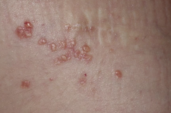
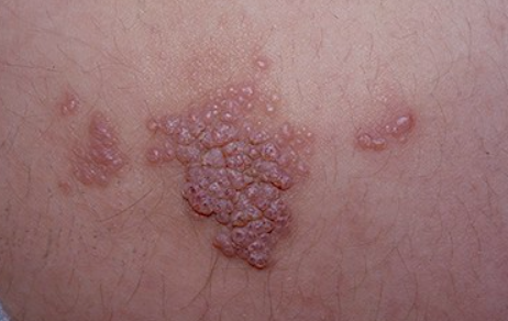
Lymphangioma - a benign tumor originating from lymphatic vessels. ICD-10 code: D18.1
Classification:
- Microcystic vascular-lymphatic malformation
- Capillary (simple, localized, lymphangioma circumscriptum, superficial microcystic malformation)
- Cavernous (cavernous lymphangioma, deep microcystic malformation)
- Macrocytic vascular-lymphatic malformation (cystic lymphangioma, cystic hygroma)
Microcystic vascular-lymphatic malformation is considered a limited anomaly of the lymphatic network during the embryonic period, resulting from vascular endothelium with lymphatic phenotype
Approximately 50% of congenital cases are detected at birth, and approximately 90% occur by 2 years of age, with an incidence of 1.2-2.8 per 100,000 newborns.
Acquired (secondary capillary lymphangiomas) develop after surgery and/or radiotherapy for malignant neoplasms at various sites.
Macrocytic vascular-lymphatic malformation begins to form in early stages of embryonic development, around the 8th to 9th week of gestation, when the lymphatic-venous system is developing.Lymphangioma circumscriptum
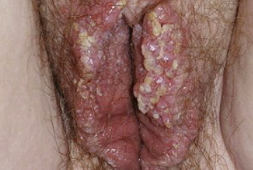
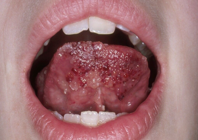
The clinical manifestations of congenital and acquired lymphangiomas are not different. They appear as small (from a few millimeters to 1-2 centimeters) raised cystic formations with the color of normal skin or reddish-brown (due to the presence of blood), grouped or arranged linearly, often against a slightly firm edematous background. The localization can be anywhere, but more commonly affects proximal parts of the limbs, neck, less commonly the genitalia, oral mucosa and periorbital area. Cases with zosteriform arrangement of elements, unilateral involvement of the abdomen, buttocks and thighs have been described. Classical lymphangiomas are usually not associated with subjective sensations.
Congenital malformations tend to progress slowly, especially during the first 2-5 years of life, with stabilization of growth occurring in the post-pubertal period. This tendency is also observed in adults with secondary lymphangiomas. Spontaneous regression is extremely rare.Vulvar lymphangioma circumscriptum
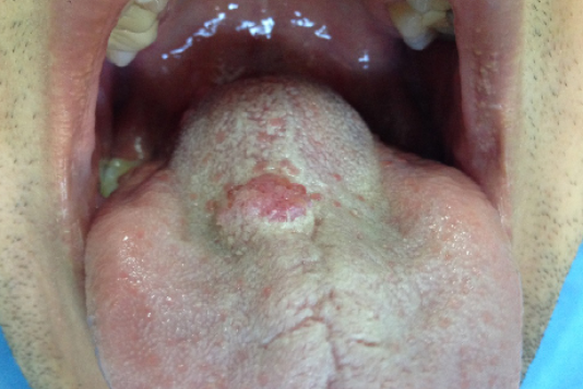
Oral Lymphangioma circumscriptum
Cavernous Lymphangioma
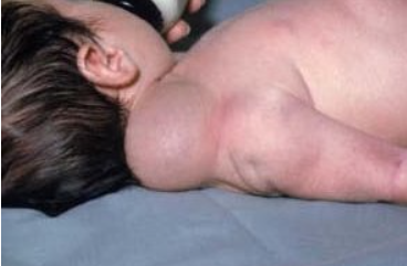
Cystic Lymphangioma
Diagnosis is based on clinical presentation and results of histological examination.
Ultrasound examination can detect cystic lymphangiomas as early as the first and second trimesters of pregnancy.- Lymphangiosarcoma
- Kaposi Sarcoma
- Angiokeratoma
- Herpes Zoster
- Morphea
- Congenital elephantiasis
- Leiomyoma
- Angiofibroma
- Warts
- Pachydermia
- Lipoma
- Infantile Hemangioma
Capillary and Cavernous Lymphangiomas
- Surgical excision to the deep fascia if a deep component is present. Recurrence occurs in 25-50% of patients within 6-80 months.
- Cryotherapy is not very effective.
- Sclerotherapy provides short-term effects and various complications.
- Laser therapy (CO2, ND:YAG)
- Radiofrequency ablation
- Oral administration of sildenafil (selective phosphodiesterase-5 inhibitor)
Cystic Lymphangiomas
- For single-chamber cysts, periodic aspiration of their contents is recommended.
- Sclerosing therapy using bleomycin, sodium tetradecyl sulfate, OK-432 (picibanil) is effective in 87-95% of cases.
- With surgical treatment, recurrences occur in 10-27% of cases with complete excision and 88% with partial removal.

