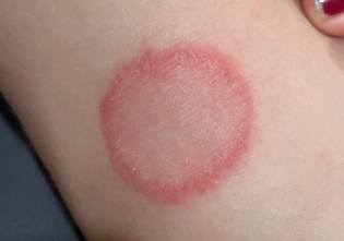
Tinea corporis (ringworm) – a superficial skin body infection caused by fungi. ICD-10 codes: B35.4
The most common causative agents of tinea corporis are the pathogenic fungi Trichophyton rubrum (90%). Less commonly, these diseases are caused by Epidermophyton floccosum and Candida species.
Infection with pathogenic fungi can occur through direct contact with an infected person, as well as through footwear, clothing, personal items (bathroom mats, sponges, nail care tools, etc.), visiting gyms, saunas, swimming pools. Skin abrasions, tears in the interdigital folds caused by friction, increased sweating or dryness of the skin, inadequate drying after water procedures, narrow interdigital spaces, vascular diseases in limbs, and other factors can facilitate fungal penetration of the skin.
Mycoses can become widespread, especially in the presence of coexisting conditions such as endocrine disorders, often diabetes, immune disorders, genodermatoses, blood disorders, and the use of antibiotics, corticosteroids, and cytostatic drugs.Tinea corporis can be caused by any dermatophyte, but most commonly by T. rubrum, less commonly by E. floccosum, T. mentagrophytes var. interdigitale, and M. canis. Risk factors include tinea pedis (caused by Trichophyton rubrum, Trichophyton mentagrophytes), contact with infected animals (Trichophyton verrucosum, Microsporum canis), and contaminated soil (Microsporum gypseum).
In zoonotic fungi, lesions are usually small and medallion-shaped. Inflammatory reactions are pronounced, often accompanied by the presence of vesicles and pustules within the lesion. In anthropophilic microsporosis caused by M. ferrugineum, lesions are larger, tend to grow peripherally, and have a "ring within a ring" appearance. Zoonotic microsporosis lesions in children are typically found on covered areas of the body, such as trunk. Key clinical features of a typical lesion of tinea corporis include:
- Clear boundaries;
- Peripheral growth;
- Ring-like shape with an inflammatory rim at the periphery;
- Resolution of inflammatory phenomena at the center of the lesion.
Tinea corporis caused by Trichophyton rubrum is often associated with tinea pedis, onychomycosis, and occasionally tinea cruris. Peripheral growth and coalescence of lesions with the formation of moderate erythema and scaling are characteristic. This condition is most commonly observed on the buttocks, thighs and shins, but can be localized on any part of the body, including the facial skin.
Different forms of tinea corporis caused by Trichophyton rubrum are erythematosquamous, follicular-papular, and infiltrative-pustular.
The erythematosquamous form is characterized by the presence of round patches of pink or reddish color, often with a bluish tinge, with well-defined borders. Small scales are usually present on the surface of the patches, and there is a discontinuous border of succulent papules around their periphery. These papules often have small vesicles and crusts.
The follicular-papular form involves the fine hairs within the erythematosquamous lesions. Hair loses its natural shine and becomes dull.
Diagnosis of dermatophytosis is based on clinical presentation and laboratory findings:
Microscopic examination of scales from affected skin areas. Cultural and PCR methods are used to determine fungal species.
If systemic antifungals are prescribed, a biochemical blood test is recommended to assess bilirubin, AST, ALT, GGT, alkaline phosphatase and glucose levels. For treatment-resistant forms of onychomycosis, ultrasound examination of superficial and deep vessels is recommended.- Pityriasis rosea
- Granuloma Annulare
- Nummular Eczema
- Psoriasis
- Necrobiosis Lipoidica
- Bowen's Disease
- Lyme Disease
- Fixed drug eruption
- Erythema annulare centrifugum
- Pityriasis Versicolor
- Contact Dermatitis
- Lichen Planus
- Seborrheic Dermatitis
- Lichen Simplex Chronicus
- Scabies
- Sarcoidosis
- Small Plaque Parapsoriasis
- Pityriasis Lichenoides
- Secondary Syphilis
- Pellagra
- Cutaneous T-cell Lymphoma
- Necrolitic Migratory Erythema
- Subacute Cutaneous Lupus Erythematosus
- Ecthyma and Impetigo
Treatment Goals:
- Clinical resolution
- Negative results in microscopy test
Topical Therapy
Topical antifungals:
- Isoconazole cream, applied 1-2 times daily for 4 weeks, or
- Terbinafine spray or dermgel, applied 2 times daily until clinical manifestations resolve, or
- Ciclopirox cream, applied 2 times daily until clinical manifestations resolve, or
- Clotrimazole cream, ointment, or solution, applied 2 times daily until clinical manifestations resolve, or
- Ketoconazole cream or ointment, applied 1-2 times daily until clinical manifestations resolve, or
- Bifonazole cream, applied 1-2 times daily for 5 weeks.
- Econazole cream, applied 2 times daily until clinical manifestations resolve, or
- Miconazole cream, applied 2 times daily until clinical manifestations resolve, or
For significant hyperkeratosis in tinea, a preliminary removal of the epidermal stratum corneum is performed using bifonazole, applied once daily for 3-4 days.
Systemic therapy
If topical therapy is ineffective, systemic antifungals are prescribed:
- Itraconazole 200 mg orally daily after meals for 7 days, then 100 mg orally daily after meals for 1-2 weeks, or
- Terbinafine 250 mg orally daily after meals for 3-4 weeks, or
- Fluconazole 150 mg orally after meals once a week for at least 3-4 weeks.
Prevention
Primary prevention: Foot care aimed at preventing microtraumas, abrasions, elimination of hyperhidrosis (aluminum chlorohydrate 15% + decylene glycol 1%) or dryness of the skin (tetranyl U 1.5% + urea 10%), flat feet, and others.
Secondary prevention: Disinfectant treatment of footwear, gloves once a month until complete healing:
Chlorhexidine bigluconate, 1% solution.
